

| Feature | What the slide or report shows | Planning use |
|---|---|---|
| Microvascular proliferation | Extensive, one of the histologic hallmarks | Look for it early; it separates glioblastoma from tumours that only look cellular and atypical. |
| Necrosis | Prominent, recognised in its pseudopalisading form | Treat as the anchor feature on H&E and sample widely, because the tumour is heterogeneous. |
| Mitoses | Frequent | Supports a high-grade reading alongside the two features above. |
| GFAP and S-100 protein | Typically positive | Use to confirm astrocytic lineage, not to assign grade. |
| WHO grade | Glioblastoma, IDH-wildtype is CNS grade 4 (2021) | Grade follows the criteria, so check the molecular side of the report as well as the slide. |
| Variants | 2 histological variants plus several other patterns, including gliosarcoma | Name the variant only when its distinguishing feature is clearly present. |
Figures above are drawn from the UPMC pathology case discussion and the National Brain Tumor Society glioblastoma overview.
Glioblastoma histology is defined by extensive microvascular proliferation, prominent necrosis and frequent mitoses in an astrocytic tumour. Confirm vascular proliferation and necrosis on H&E first, then use variant features and molecular findings to settle the rest.
This guide follows the order in which a slide is read, from gross context to molecular criteria. For the wider classification framework, see our overview of brain tumor pathology.
Gross context and diagnostic setting
Glioblastoma, IDH-wildtype is considered grade 4 by the WHO, and the diagnostic criteria changed in the 2021 classification, according to the National Brain Tumor Society. It belongs to the glioma group, specifically the astrocytoma subtype, as MD Anderson describes it.
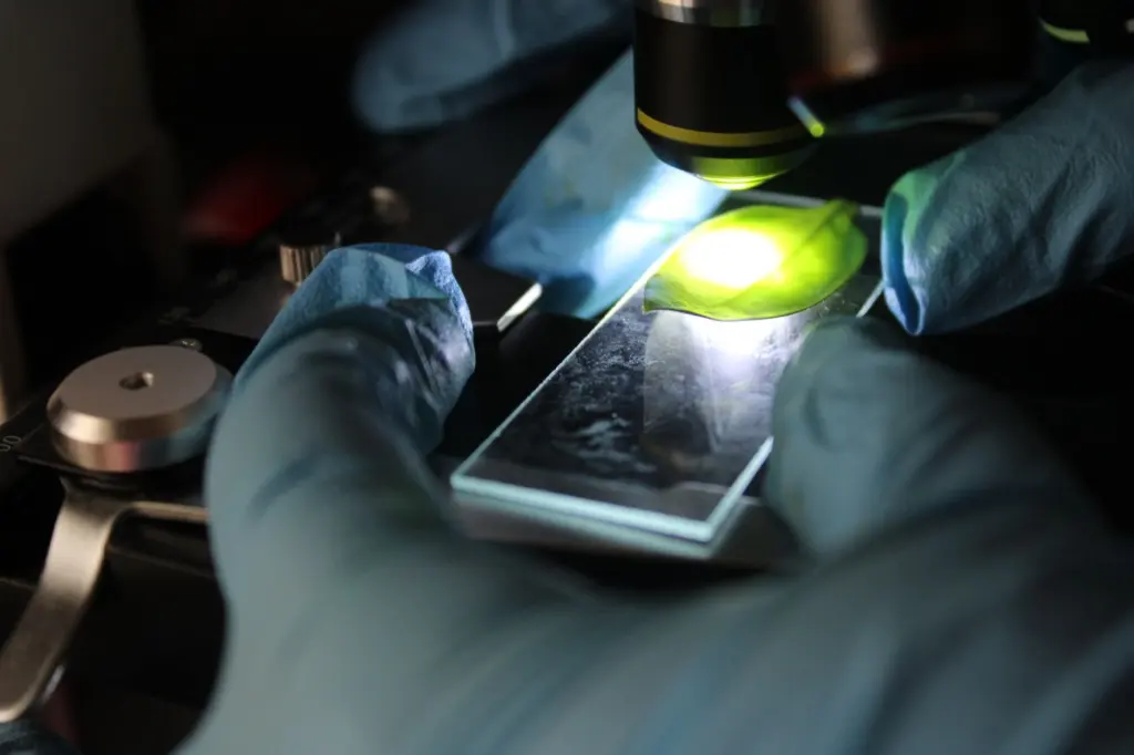
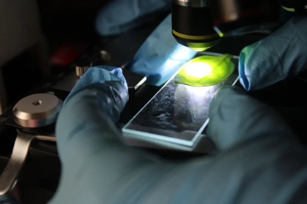
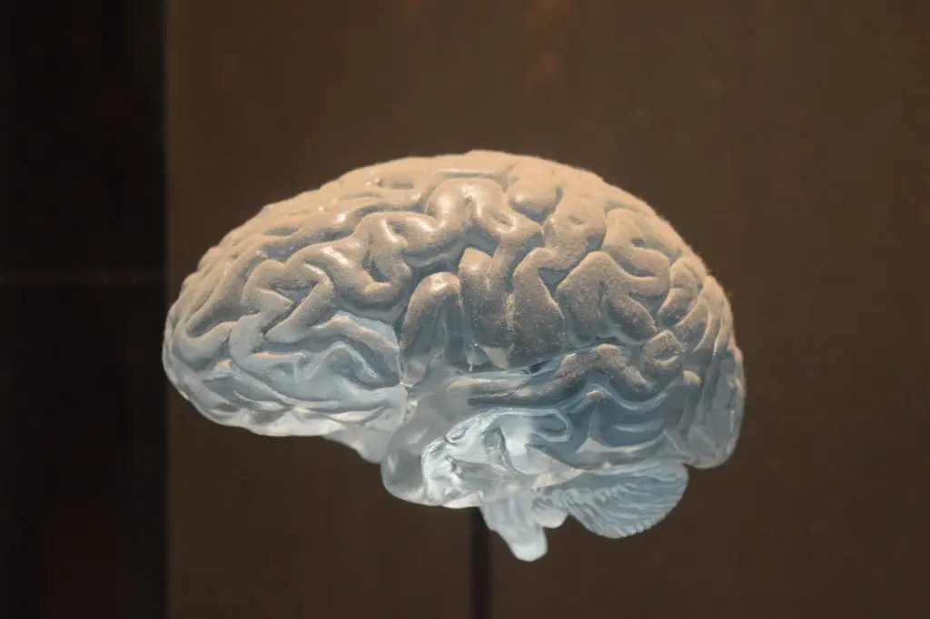
Most cases can be definitively diagnosed after surgery through histological testing, when a neuropathologist examines tissue under a microscope. The slide is therefore the decision point. Gross appearance only sets expectations for what the sections should show.
Reading the H&E slide
Glioblastoma is morphologically heterogeneous, with extensive microvascular proliferation, prominent necrosis and frequent mitoses as its histological hallmarks, according to the UPMC case discussion. Because the appearance varies from field to field, a single region lacking one hallmark does not settle the diagnosis. Work through the sections in a fixed order.
- Scan for necrosis. Look for prominent necrosis at low power and note whether it has the pseudopalisading form.
- Look for vascular change. Search for extensive microvascular proliferation in cellular areas and beside necrosis.
- Assess mitoses. Frequent mitoses support a high-grade reading.
- Note pleomorphism. Record any cellular pleomorphism and multinucleated cells, since these point towards a variant.
Pseudopalisading necrosis
Pseudopalisading necrosis is the pattern of necrosis this guide treats as the defining slide feature of glioblastoma. It is the first thing to confirm on a suspicious section. Where necrosis is present but its pattern is unclear, sample more widely before committing to a reading, since the tumour is heterogeneous.
Mitoses and pleomorphism
Frequent mitoses and marked pleomorphism show how aggressive the tumour is, but they do not define glioblastoma alone. Use them as supporting evidence once necrosis or vascular proliferation has been found.
Microvascular proliferation
Microvascular proliferation is the overgrowth of endothelial cells in the tumour’s blood vessels, and it is a core histologic hallmark of glioblastoma. In the UPMC case, endothelial proliferation appears together with cellular pleomorphism and multinucleated cells in a malignant tumour.
It matters in two ways. Diagnostically, extensive vascular proliferation is one of the features that places a tumour in the glioblastoma group. Therapeutically, the tumour’s vessels are a treatment target: bevacizumab inhibits the growth of blood vessels that supply the tumour, as described by the Global Coalition for Adaptive Research.
GFAP and glial lineage
As an astrocytic neoplasm, glioblastoma typically stains positively for GFAP and S-100 protein, per the UPMC discussion. GFAP staining therefore confirms glial differentiation in the tumour cells.
Use GFAP to answer the lineage question, not the grade question. Grade comes from necrosis and vascular proliferation on H&E, together with the molecular findings covered below.
Histologic variants and gliosarcoma
In addition to the classic form, 2 histological variants and several other patterns are defined, including gliosarcoma, according to the UPMC case discussion. The same source names giant cell glioblastoma, glioblastoma with lipidized cells and small cell glioblastoma. It also lists patterns with an oligodendroglioma component and heterologous differentiation.
| Pattern | Distinguishing feature | What to check |
|---|---|---|
| Classic glioblastoma | Microvascular proliferation and necrosis without a dominant additional feature | Confirm both hallmarks before looking for a variant. |
| Gliosarcoma | Biphasic pattern of glial and spindle cell areas | Examine the spindle cell areas alongside the glial areas; a reticulin stain helps show the pattern. |
| Giant cell glioblastoma | Bizarre multinucleated cells with pleomorphism | Judge whether the multinucleated cells dominate the tumour. |
| Other named patterns | Lipidized cells, small cells, an oligodendroglioma component or heterologous differentiation | Record the pattern as a descriptive feature on top of the hallmarks. |
Pattern names are taken from the UPMC pathology case discussion.
Gliosarcoma pathology
Gliosarcoma is the variant to think of when a glioblastoma contains two distinct tissue components. The slide shows glial areas next to spindle cell areas, and the reticulin stain makes the contrast between them visible. The glial component keeps the usual hallmarks; the spindle component is what sets this variant apart.
Giant cell glioblastoma
Giant cell glioblastoma is recognised by bizarre multinucleated cells within a pleomorphic tumour. The UPMC case describes cellular pleomorphism with multinucleated cells in a malignant tumour. Multinucleated cells alone do not make this variant. It is the variant to name when they dominate the field, because pleomorphism also occurs in classic glioblastoma.
Molecular criteria and the 2021 WHO changes
The 5th edition of the CNS WHO classification introduced several major changes in practice, including molecular grading of adult diffuse gliomas, as the Weill Cornell record of the review notes. The National Brain Tumor Society likewise states that the diagnostic criteria for glioblastoma changed in 2021.
The practical consequence is that histology is no longer the whole diagnosis. A tumour that shows neither prominent necrosis nor vascular proliferation on the slide is not automatically excluded from glioblastoma, IDH-wildtype once molecular findings are applied. Read the molecular section of the report as part of the diagnosis, and check the exact marker list against the WHO 5th edition text.
Troubleshooting a borderline biopsy
- No necrosis in the sample. Heterogeneity means a small sample may miss it. Look for vascular proliferation, and rely on the molecular findings for diagnostic criteria that do not need necrosis.
- Spindle cell areas. Consider gliosarcoma and look for the biphasic glial and spindle pattern, using reticulin if needed.
- Bizarre multinucleated cells. Consider giant cell glioblastoma, but confirm the cells dominate the tumour before naming the variant.
- Hallmarks absent and tumour looks low grade. Compare against a different entity such as the one in our guide to pilocytic astrocytoma histology.
- Unexpected stain results. GFAP and S-100 are typically positive in glioblastoma, so a negative result should prompt a review of lineage.
Frequently asked questions
Is glioblastoma diagnosed from histology alone?
Most cases can be definitively diagnosed after surgery through histological testing, according to MD Anderson.
Does a positive GFAP stain prove glioblastoma?
No. GFAP positivity shows astrocytic differentiation, which glioblastoma typically has. Grade rests on necrosis and vascular proliferation and on the molecular findings.
How common is glioblastoma, IDH-wildtype?
Under the prior criteria, it made up 13.9% of all brain and other CNS tumours, with a median age at diagnosis of 66 years, as reported by the National Brain Tumor Society for 2017-2021.
Why is gliosarcoma treated as a glioblastoma pattern?
The UPMC case discussion lists gliosarcoma among the histological variants and patterns defined in addition to classic glioblastoma. Its spindle cell component distinguishes it from the classic form.
Leave a Reply