
| Feature | What to look for | Planning use |
|---|---|---|
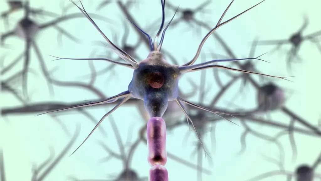
| Neuron | Cell body (perikaryon), axon and dendrites; cell body up to 135 µm in motor neurons, 4 µm in cerebellar granular neurons | Judge size first. Motor neurons are the largest cells in the CNS. |
| Glial cells | Astrocytes, oligodendroglia, ependymal cells and microglia | Once a neuron is excluded, sort what remains into these four types. |
| Nissl substance | Aggregates of rough endoplasmic reticulum, visible with Nissl stain and H&E | Treat it as the baseline against which chromatolysis is judged. |
| Red neuron | Shrunken cell, eosinophilic cytoplasm, pyknotic nucleus | Read it as irreversible injury, for example from hypoxia, ischemia or hypoglycemia. |
| Central chromatolysis | Ballooned cell body, disaggregated Nissl granules, smooth cytoplasm, peripheral nucleus | Read it as a reversible repair response after axon transection. |
| Neurofibrillary tangle | Insoluble hyperphosphorylated tau in the perikaryon, built of paired helical filaments | Associated with Alzheimer disease and other tauopathies; confirm with silver stain. |
These values come from the Neuropathology Web chapter on neurons and glia, which describes the normal cells and their basic reactions to injury.
Neurocytology starts with telling a neuron from a glial cell on H&E, then asking whether the neuron looks normal, acutely injured, reacting to axonal damage or carrying an inclusion.
This page is the cell-level foundation for the neuropathology course. Later disease topics assume you can already recognise these patterns, and the glial cells are developed further in astrocytes in neuropathology.
Start with the normal neuron
The central nervous system consists of neurons and glial cells. Every neuron has a cell body, an axon and dendrites. Dendrites receive signals and the axon conveys them onward.
Size is the quickest first discriminator. Motor neurons are the largest cells in the CNS, with a cell body up to 135 µm. Cerebellar granular neurons measure about 4 µm. A section can therefore contain neurons that look very different, and no single “typical neuron” appearance applies across the brain.
Internal structure that matters at the microscope
The cytoplasm contains Nissl granules, which are aggregates of rough endoplasmic reticulum. They are easy to see with the Nissl stain and with H&E, so a normal neuron has a textured, granular cytoplasm. Remember this appearance, because both injury patterns below change it.
Neurofilaments are 10 nm intermediate filaments, and neurotubules measure 20 to 26 nm. Neither is resolved on routine light microscopy. They matter because immunohistochemistry can demonstrate cytoskeletal proteins, and synaptophysin in synaptic vesicles, when a stain needs to confirm that a cell is a neuron.
Neuropil and function
The fibrillary “matrix” of the cerebral gray matter is the neuropil. It is formed by the cellular processes of neurons and glia with minimal extracellular space. When you see pale, finely fibrillar background between cell bodies in gray matter, you are looking at these tangled processes rather than at empty space.
Functionally, synaptic vesicles contain synaptophysin and synapsins. Glutamate is the most important excitatory neurotransmitter, and gamma amino butyric acid (GABA) is the most important inhibitory one.
Sort the glia
The glial cells are astrocytes, oligodendroglia, ependymal cells and microglia. Astrocytes have a clear anatomical relationship you can use: in brain capillaries, they are apposed to the vessel wall. A glial nucleus sitting beside a capillary is therefore a reasonable place to look for astrocytes.
- Exclude neurons first. Look for a cell body with a recognisable perikaryon and granular Nissl cytoplasm.
- Look at the remaining nuclei. Note where they lie relative to vessels, neuropil and neuronal cell bodies.
- Assign the glial type. Choose among astrocyte, oligodendroglial cell, ependymal cell and microglial cell, and confirm uncertain cases with a stain rather than guessing from H&E alone.
Each glial type has its own injury responses and tumour counterparts, which later course topics develop. Astrocytic reactions in particular are covered on their own page, astrocytes in neuropathology.
Choose the stain
H&E is adequate for routine study of cellular details of neurons and glia. It does not stain neuronal processes, so it will not show you axons or dendrites in detail. Choose H&E for cell body shape, nuclear change and cytoplasmic colour, and move to another method when the question concerns processes or specific proteins.
- H&E. The routine starting point for cell bodies, nuclei and eosinophilia.
- Nissl stain. Makes Nissl granules easy to see, which is useful when judging chromatolysis.
- Bielschowsky silver stain. The most commonly used silver stain for axons and dendrites, and it also shows Alzheimer lesions.
- Immunohistochemistry. Demonstrates cytoskeletal proteins and synaptophysin in nerve cells.
If tissue is sampled poorly, no stain rescues it. How blocks are taken is covered in the guide to neuropathology brain dissection.
Recognise acute injury: red neurons
Injured neurons shrink, become eosinophilic because mitochondria condense, and develop pyknotic nuclei. This shrunken, eosinophilic appearance is what is meant by a red neuron. On H&E the cytoplasm turns a uniform pink-red and loses its granular texture.
Red neurons signal irreversible injury. Hypoxia, ischemia and hypoglycemia cause irreversible neuronal injury, so finding these cells in a section points you toward one of those insults rather than toward a reversible change. They do not by themselves identify which insult occurred, so interpret them with the clinical history and the distribution of the change.
- Check cell size. The cell body should look shrunken compared with neighbouring neurons.
- Check the cytoplasm. Look for homogeneous eosinophilia in place of visible Nissl granules.
- Check the nucleus. Pyknosis supports acute injury.
Recognise the axonal reaction: central chromatolysis
Central chromatolysis is a reversible change that develops during repair of a neuron disconnected from its target. It occurs after axon transection. If the axon is transected, the rough endoplasmic reticulum disaggregates and the neuronal body balloons.
The features are a swollen cell body, dispersed Nissl granules, smooth cytoplasm and a nucleus displaced to the periphery. Compare this with a red neuron: central chromatolysis is a swollen, pale-centred cell, while a red neuron is shrunken and dense. Because one pattern is reversible and the other is not, confusing them changes the interpretation completely.
Use a Nissl stain or H&E to confirm. The loss of the normal granular Nissl substance in the central cytoplasm is the defining feature, so it helps to have a normal neuron in the same field for comparison.
Inclusions in neurons
Neuronal inclusions are abnormal protein deposits within the cell. The clearest example in this reference is the neurofibrillary tangle: insoluble, hyperphosphorylated tau deposited in the neuronal perikaryon in Alzheimer disease and other tauopathies, formed of paired helical filaments.
The Bielschowsky silver stain is the practical way to demonstrate such lesions in tissue, because H&E does not highlight them as well. Immunohistochemistry adds protein-specific confirmation. The wider range of inclusions and the diseases that produce them are developed in the neurodegenerative disease topics that build on this page.
Reading further
For a full textbook treatment, Ennio Pannese’s Neurocytology was published by Thieme in 1994 and runs to 264 pages.
Frequently asked questions
Which stain shows axons and dendrites?
The Bielschowsky silver stain is the most commonly used silver method for them. H&E does not stain neuronal processes.
Can a red neuron recover?
No. Red neurons reflect irreversible injury, such as that from hypoxia, ischemia or hypoglycemia. Central chromatolysis, by contrast, is reversible.
What is the neuropil?
It is the fibrillary matrix of cerebral gray matter, made of the processes of neurons and glia with minimal extracellular space.
Which glial cells lie against capillaries?
Astrocytes. In brain capillaries they are apposed to the vessel wall.
Leave a Reply