
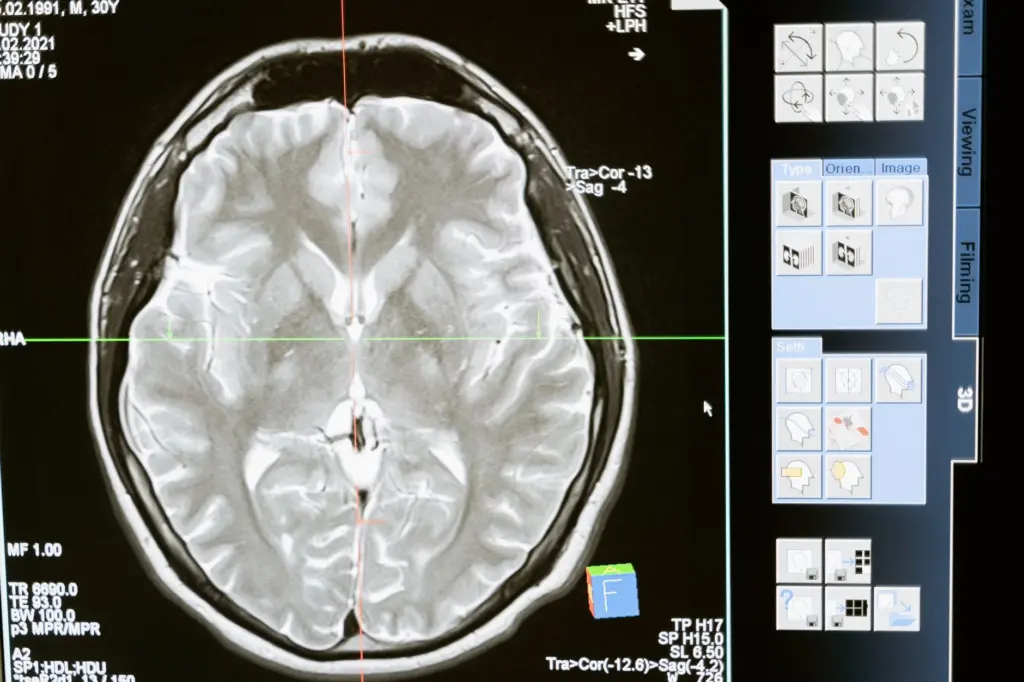
Brain tumor pathology begins with one decision: decide whether the tumor started in the brain or spread to it from elsewhere. Then classify by histology, molecular markers and within-type WHO grade, starting with IDH and 1p/19q status for adult gliomas.
This page teaches the sorting logic. For a full catalogue of entities, use the guide to types of brain tumors, then return here when you need to decide which feature to check first.
Choosing where to start
The right starting point depends on what you already know about the specimen or the case. Pick the branch that matches, and read the section it points to before the others.
- The tumor may have come from another organ. Metastatic cancer is the most common type of brain cancer according to Northwestern Medicine, so rule it in or out first. Go to the primary versus metastatic section.
- The tumor is an adult diffuse glioma. Start with IDH mutation and 1p/19q codeletion status, because those two results sort adult-type diffuse gliomas into three types. Go to the diffuse glioma section.
- The tumor is a high-grade glioma in the midline, especially in a young patient. Check for an H3 K27 alteration before applying the adult glioma logic.
- The tumor is a small-cell embryonal tumor of the cerebellum. Think medulloblastoma and move straight to the molecular group question.
- The tumor shows whorls, palisades or finger-like fronds. Think meningioma, schwannoma or choroid plexus papilloma, and read the section on meningeal, nerve-sheath and other tumors.
Whichever branch you choose, three questions run underneath it. What does the tissue look like, which molecular marker defines or refines the entity, and what grade applies within that entity? A diagnosis that answers all three is stronger than one that answers only the first. A student who memorizes appearance alone will be caught out by tumors that look alike but differ in their markers, and a student who memorizes markers alone will not know what to look for on the slide.

If you are comparing the broad term with its most aggressive member, the explainer on glioma vs glioblastoma is the quickest route to that distinction. The sections below keep the decision logic in one place.
How the WHO classification is organized
The WHO classification of central nervous system tumors is the shared vocabulary of the field. It assigns each tumor a type, a molecular or histologic definition and a grade, and it changes with each edition. Treat the edition you are reading as part of the diagnosis.
From microscopy alone to integrated diagnosis
Older diagnosis relied on the microscope. A review in PubMed Central notes that review panels revealed considerable diagnostic inter-observer variation in microscopy-based CNS tumor diagnosis. The WHO 2016 update responded by making molecular aberrations part of the definition of particular brain tumor entities for the first time.
This is why a modern tumor name often contains a gene. The name is not decoration. A tumor called astrocytoma, IDH-mutant or oligodendroglioma, IDH-mutant and 1p/19q-codeleted cannot be assigned from the slide alone, because the marker is written into the definition. For a learner, the practical consequence is that the diagnostic card for each tumor must have a molecular line, not only a histology line.
The same review lists the defining molecular markers for diffuse gliomas: IDH1/IDH2 mutations, 1p/19q codeletion and mutations in histone H3 genes. Those three families recur throughout the glioma sections below, and they are the best place to anchor your memory before you add entity-specific detail.
Grades are assigned within tumor types
The fifth edition of the WHO classification was released in 2021, and a review in the Journal of Pathology and Translational Medicine describes two changes that affect how you read a diagnosis. CNS WHO grades, previously written in Roman numerals I to IV, are now written in Arabic numerals 1 to 4. Grading is also done within tumor types rather than across types.
The second change matters more than the first. Within a type, the grade carries the information, and astrocytoma, IDH-mutant is assigned grade 2, 3 or 4. Oligodendroglioma, IDH-mutant and 1p/19q-codeleted is assigned grade 2 to 3. A grade 3 in one type is therefore not a statement about a grade 3 in another type. When you compare tumors, compare their types first and their grades second.
Older teaching material and older reports use Roman numerals and the earlier naming. If you meet a diagnosis written that way, read it as belonging to the earlier scheme and translate it carefully, not by simple substitution of numerals.
Primary and metastatic brain tumors
A primary brain tumor starts in the brain, and a metastatic tumor spreads to the brain from another part of the body. Northwestern Medicine states that metastatic cancer is the most common type of brain cancer, which is why it belongs at the top of any decision path.
The distinction changes everything that follows. A primary tumor is classified using the WHO scheme described above, with its type, markers and grade. A metastasis is named for the cancer it came from, so the pathologist’s job is to identify the origin.
What metastases look like
The same Northwestern Medicine article explains that neuropathologists can determine a tumor’s origin from its cell patterns and from special tests. Two examples are useful for learners. Metastatic cancer from the colon tends to form little glands within the brain, and these glands are filled with debris. Metastases from the breast or lung can produce bluish-grey mucus.
Those are pattern cues, not a complete list. Use them as the start of a differential, and expect to confirm the origin with special tests. A glandular or mucinous pattern in brain tissue should make you ask where a gland-forming cancer might have started, rather than force the tissue into a glioma category.

Using the distinction in practice
Ask the origin question before the grade question. A primary diffuse glioma earns its grade within its type, while a metastasis is described by where it came from. Mixing the two frameworks is a common beginner error, and it is easy to avoid if you keep the primary-or-metastatic decision first.
Diffuse gliomas: marker-first recognition
For adult-type diffuse gliomas, the molecular result comes before the histologic label. The Journal of Pathology and Translational Medicine review explains that these tumors are classified as one of three types, based on the presence or absence of IDH mutation and 1p/19q codeletion. The decision path is short enough to hold in your head.
- IDH-wildtype, with the right features: glioblastoma, IDH-wildtype.
- IDH-mutant, no 1p/19q codeletion: astrocytoma, IDH-mutant.
- IDH-mutant with 1p/19q codeletion: oligodendroglioma, IDH-mutant and 1p/19q-codeleted.
Learn this path as a flowchart first and attach histology afterward. It will save you from the trap of guessing the type from appearance and then having a marker contradict you.
Glioblastoma, IDH-wildtype
Glioblastoma is the most common and most malignant tumor that starts within the brain itself, according to Northwestern Medicine. The WHO fifth edition reserves the term specifically for IDH-wildtype adult-type diffuse gliomas, so an IDH-mutant glioma is no longer called glioblastoma.
The diagnostic criteria require the absence of IDH and H3 mutations, together with the presence of either microvascular proliferation or tumor cell necrosis. Alternatively, the diagnosis can rest on TERT, EGFR or +7/-10 markers. The histologic features and the molecular features are therefore alternative routes to the same diagnosis in an IDH-wildtype tumor, and a pathologist can use either.

For the slide-level details, the dedicated glioblastoma histology page expands on what to look for. When the question is how this tumor relates to the broader glioma group, remember that the term glioblastoma now carries a marker requirement, not only a histologic one.
Astrocytoma, IDH-mutant
Astrocytoma, IDH-mutant is the entity that most clearly shows the within-type grading rule. It is assigned grade 2, 3 or 4, so the same name spans a range of behavior. The type tells you the lineage and the marker, and the grade tells you where in that range the individual tumor sits.
The practical rule for students is simple. When you see IDH mutation and no 1p/19q codeletion in a diffuse glioma, the type is astrocytoma, and the grade must still be assigned. An IDH-mutant tumor that reaches the highest grade stays an astrocytoma and is not relabeled glioblastoma. The page on astrocytoma histology covers the grading features in depth.
Pilocytic astrocytoma has its own page. Read pilocytic astrocytoma histology as a comparison with the IDH-mutant astrocytoma above, and keep the two entities separate in your notes so that a shared word in the name does not blur their definitions.
Oligodendroglioma, IDH-mutant and 1p/19q-codeleted
Oligodendroglioma is the entity with the strictest molecular definition. Diagnosis requires an IDH1 or IDH2 mutation and a concurrent 1p/19q codeletion, and the tumor is assigned CNS WHO grade 2 to 3. If either marker is missing, the diagnosis is not oligodendroglioma.
This is the tumor where a purely histologic habit does the most harm. A tumor that resembles an oligodendroglioma on the slide but lacks the codeletion must be classified by its molecular result. The oligodendroglioma histology guide pairs the microscopic picture with this requirement.
Diffuse midline glioma, H3 K27-altered
Pediatric-type diffuse high-grade gliomas form a separate branch, and they include diffuse midline glioma, H3 K27-altered. The classification describes it as a CNS WHO grade 4 diffusely infiltrating glioma. The defining marker here is the histone alteration, which is the third of the glioma marker families after IDH and 1p/19q.
The lesson for the decision path is that H3 status is a separate question from IDH status. A high-grade glioma in a young patient should prompt you to look for the histone alteration, not to assume the adult flowchart applies. For wider growth-pattern questions in diffuse tumors, the page on gliomatosis cerebri pathology is a useful companion.
Embryonal and ependymal tumors
These tumors do not follow the IDH and 1p/19q path, so treat them as their own branch. Their classification leans on molecular subgrouping within a named entity.
Medulloblastoma
Medulloblastomas are the most frequent embryonal CNS tumors, and the 2016 WHO classification divided them into four molecularly defined groups, as the PubMed Central review describes. The groups are WNT, SHH TP53-mutant, SHH TP53-wildtype, and non-WNT/non-SHH.
Notice that the SHH group is split by TP53 status, while the last group is defined negatively as neither WNT nor SHH. When you write your own summary, copy the group names exactly, because small changes in wording change the meaning. The page on medulloblastoma histology connects the groups to the microscopic picture.
Ependymoma
Ependymoma is the other entity in this branch where a molecular subtype appears in the diagnosis. The same review identifies the RELA fusion-positive subtype of ependymoma as a molecular diagnostic feature. For a learner, the takeaway is that a morphologic diagnosis of ependymoma can be refined by a fusion result.
Use the page on ependymoma histology for the slide-level appearance, and keep the RELA fusion in your notes beside the name so that the molecular line is never left blank.
Meningeal, nerve-sheath and other tumors
Several common tumors are recognized mostly by pattern, and these are the best entities for building microscope confidence. Northwestern Medicine describes distinctive features for each, and the descriptions work well as quick recognition cues.
- Meningioma: characterized by distinctive pink whorls of cells. The whorl is the cue that most quickly points a student to the diagnosis.
- Schwannoma: a benign tumor with cells forming neat palisades, nearly always noncancerous. Read the dedicated schwannoma histology page for the full picture.
- Choroid plexus papilloma: presents as finger-like structures and is typically low-grade. See choroid plexus papilloma histology for more.
- Chordoma: a rare tumor with its own diagnostic page, chordoma pathology.
These tumors reward visual learning. The cue words are whorls, palisades and fronds, and each is a pattern you can check at the microscope without a molecular test. Pair each pattern with its entity name in a flash card and test yourself on images, not only on text.
Some further entities have pages of their own: craniopharyngioma histology, ganglioglioma histology and hemangioblastoma histology. Add them to your sorting scheme once the main branches feel secure, and not before.
Reading histology: what the microscope settles
The microscope still does a great deal of the work, but it rarely does all of it. Pattern recognition tells you which branch of the decision path you are on, and molecular results confirm or change the final name.
The documented inter-observer variation in microscopy-based diagnosis is a reason to be humble about appearance. Two careful observers can read the same slide differently. That variability is a practical argument for treating each histologic cue as a prompt for a test, not as the answer. A slide that looks like glioblastoma still needs the marker or histologic criteria listed above, and a slide that looks like oligodendroglioma still needs the codeletion.
Use the following habit when studying slides. Name the pattern you see, state the entity it suggests, then write down which marker or grade criterion would confirm or overturn it. That three-step note keeps you from treating a resemblance as a diagnosis, and it mirrors how the classification itself is built.
From scan to tissue diagnosis
Imaging shows where a lesion sits, but the classification described on this page rests on tissue. Northwestern Medicine notes that neuropathologists can usually tell from a biopsy whether a tumor is low-grade or malignant, what kind of cancer is present and what its grade is.
For a learner, the workflow means that each diagnosis is the product of several steps. The scan places the lesion, the tissue shows the pattern, special tests and molecular results supply the markers, and the WHO edition supplies the name and the grade. Skipping any step leaves a gap that the classification is designed to close.
Overview of recognition cues
The table below collects the diagnostic cue, grade information and marker for the main entities discussed above, so you can scan across entities on common criteria. Medulloblastoma and ependymoma are covered in their own section because their molecular subgrouping does not fit a single table cell.
| Tumor | Recognition cue | Grade or behavior | Marker or test |
|---|---|---|---|
| Glioblastoma, IDH-wildtype | Microvascular proliferation or tumor cell necrosis | Most malignant tumor starting within the brain | IDH-wildtype, no H3 mutation; or TERT, EGFR or +7/-10 |
| Astrocytoma, IDH-mutant | Diffuse glioma with IDH mutation | Grade 2, 3 or 4 within type | IDH1/IDH2 mutation, no 1p/19q codeletion |
| Oligodendroglioma, IDH-mutant and 1p/19q-codeleted | Diffuse glioma defined by both markers | CNS WHO grade 2-3 | IDH1/IDH2 mutation plus 1p/19q codeletion |
| Diffuse midline glioma, H3 K27-altered | Diffusely infiltrating glioma | CNS WHO grade 4 | H3 K27 alteration |
| Meningioma | Pink whorls of cells | Roughly 90% low-grade | Pattern recognition |
| Schwannoma | Cells in neat palisades | Nearly always noncancerous | Pattern recognition |
| Choroid plexus papilloma | Finger-like structures | Typically low-grade | Pattern recognition |
| Brain metastasis | Glands (colon) or bluish-grey mucus (breast, lung) | Spread from another part of the body | Special tests to identify origin |
The glioma rows draw on the WHO fifth-edition review, and the pattern-based rows draw on Northwestern Medicine’s overview of brain tumors under the microscope.
A study plan built on the decision path
The most efficient way to learn brain tumor pathology is to follow the same order the diagnosis follows. Start with the coarse split, then add markers, then grades, then appearance.
- Learn the glioma flowchart first. It has three outcomes, and it organizes the largest group of entities. Once it is automatic, glioblastoma, astrocytoma and oligodendroglioma stop blurring together.
- Attach one cue to each entity. Necrosis or microvascular proliferation for glioblastoma, whorls for meningioma, palisades for schwannoma, fronds for choroid plexus papilloma. One strong cue is better recalled than a paragraph of description.
- Keep marker names exact. TP53 status splits the SHH medulloblastoma group, and the codeletion is required for oligodendroglioma. Small wording differences change the diagnosis.
- Study grades within types. Write the grade range beside each type, and never compare a grade across types.
- Revisit the primary or metastatic question for every case. It is the first branch of the path, and it is the one most often skipped when the tissue looks like a glioma.
If time is short, prioritize the glioma flowchart and the pattern-recognition tumors, because they cover the entities you are most likely to meet. Add the molecular groups of medulloblastoma and the ependymoma subtype once the basics hold.
Frequently asked questions
How many people are diagnosed with glioblastoma each year?
Northwestern Medicine states that more than 12,000 people are diagnosed with glioblastoma in the United States every year, and describes it as the most common malignant tumor starting within the brain.
Can a biopsy show whether a brain tumor is low-grade or malignant?
Usually, yes. Neuropathologists can generally tell from tissue whether a tumor is low-grade or malignant, what kind of cancer it is and what its grade is, using cell patterns and special tests.
Does a low-grade meningioma always stay controlled?
No. Roughly 90% of meningiomas are low-grade, but many grow back, and some may become resistant to radiation, according to Northwestern Medicine.
How rare is chordoma?
Chordomas occur in about one in one million people. They often keep growing back after resection and radiation, which is why they have their own dedicated pathology page.
Why are Roman numeral grades no longer used in WHO reports?
The fifth edition, released in 2021, writes CNS WHO grades in Arabic numerals 1 to 4 instead of the earlier I to IV, and it assigns grades within tumor types.
Leave a Reply