
A posterior fossa tumor shows rosette-forming cells on the slide. Ependymoma histology supports the diagnosis, but H3 K27me3 staining and tumor site decide whether it is group A or group B.
- Slide first: rosettes and perivascular pseudorosettes point to ependymoma but do not name the molecular type.
- Site second: supratentorial, posterior fossa and spinal tumors each carry their own marker sets.
- Stain third: in the posterior fossa, global loss of H3 K27me3 points to group A and retained staining to group B.
This guide sits within our wider coverage of brain tumor pathology. It works through the practical problems a reader meets: recognizing the tumor, choosing the molecular type by location, separating group A from group B, and understanding how far grading can be trusted.
Recognizing ependymoma on the slide
Ependymoma is recognized on hematoxylin and eosin sections by its ependymal differentiation, and the diagnosis is built from more than the microscope image. A neuropathology source describing current practice states that diagnosis is based not only on how the tumor looks under the microscope, but also on site and molecular features.
Rosettes and supportive stains
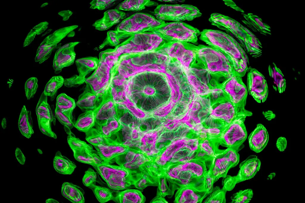
The classic feature is the perivascular pseudorosette, in which tumor cells arrange themselves around blood vessels with a cell-free zone between them. True ependymal rosettes are less common. They surround a central lumen, which separates them from pseudorosettes. Dot-like epithelial membrane antigen (EMA) staining is a useful supportive immunostain, because it highlights the ependymal character of the cells without settling the molecular type.
Gross appearance
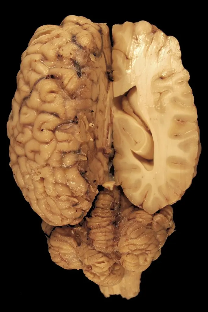
At the bench, the American Brain Tumor Association describes ependymomas as soft, grayish or red tumors that may contain cysts or mineral calcifications. In the posterior fossa the mass often sits in or near the fourth ventricle. The gross picture is a clue to site, not a substitute for microscopy.
Related tumors to compare
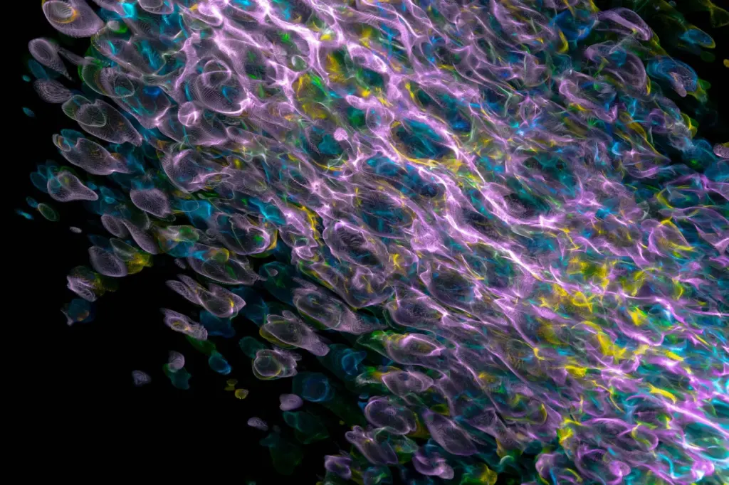
When the differential reaches into cerebral or cerebellar territory, compare the features on our pages for oligodendroglioma histology and medulloblastoma histology. Each is a separate diagnosis with its own molecular logic.

Choosing the molecular type by location
Site is the first branch point for molecular classification, because ependymomas in different compartments are defined by different markers. The table summarizes what each location tests for and how to use it.
| Site | Types and markers | Planning use |
|---|---|---|
| Supratentorial | ZFTA, RELA, YAP1, MAML2; ZFTA fusion-positive and YAP1 fusion-positive subtypes | Request fusion testing so the tumor can be assigned to a fusion-defined subtype |
| Posterior fossa | PF-A, PF-B and posterior fossa ependymoma NOS; H3 K27me3 and EZHIP (methylome) | Start with H3 K27me3 immunohistochemistry, then use methylome data where available |
| Spinal cord | NF2, MYCN | Use spinal markers rather than the supratentorial or posterior fossa panels |
The marker lists come from the American Brain Tumor Association, and the 2021 WHO posterior fossa categories are summarized by Medscape.
Why age and site travel together
Location correlates strongly with age. Medscape reports that 90% of childhood ependymomas are intracranial, while 50-60% of adult ependymomas arise in the spine. The ABTA gives a median age at diagnosis of 5 years in children and 30-40 years in adults. A tumor in a young child therefore raises intracranial types first, and a spinal mass in an adult raises the spinal marker set first.
Supratentorial tumors and ZFTA fusion
In the cerebral hemispheres, ZFTA fusion is the headline alteration, with a YAP1 fusion-positive type recognized alongside it. A routine slide cannot show a fusion, so a supratentorial tumor with ependymoma histology needs molecular confirmation before it is placed in a subtype.
Posterior fossa group A versus group B
Groups A and B are not separable by their histologic appearance. A study from Johns Hopkins describes them as morphologically identical, but biologically distinct tumor entities. That is why the stain matters more than the rosettes at this step.
What H3 K27me3 shows
H3 K27me3 staining is globally reduced in group A tumors compared with group B. Scoring is simple: the study assessed the stain as positive or negative, in a blinded manner. In a series of 230 childhood ependymomas, 112 had methylation subgroup data (group A n=72, group B n=40). Against that methylation reference, immunohistochemistry showed 99% sensitivity and 100% specificity in separating the two groups. The authors also found that the stain predicted outcome in two independent, non-overlapping cohorts (n=133 and n=97).
Age and outlook
Group A is seen mainly in younger children, and group B more often in older children and adults, according to Atlantic Brain and Spine. The Johns Hopkins authors state that group A tumors have a poor prognosis and require intensive therapy, whereas group B tumors have an excellent prognosis. This gap is the main reason accurate grouping matters, since two tumors that look the same under the microscope can call for very different management.
Why the stain is the practical route
Methylation profiling defines the groups, but the same study notes that these assays are not routinely available. A validated H3 K27me3 stain therefore gives many laboratories a workable way to assign a group.
Grading and where it is uncertain
A tumor grade describes how normal or abnormal tumor cells appear under a microscope, as the National Brain Tumor Society explains. For ependymoma the practical question is how much weight that appearance deserves.
Grade 2 versus grade 3
Medscape notes that ependymomas may receive a grade of 2 or 3 depending on histology and molecular analysis, and that the distinction between grade 2 and 3 has been controversial. Treat a histologic grade as one input rather than a stand-alone prognostic label. Two pathologists can reasonably weigh the same features differently, so it makes sense to read the grade next to the site and molecular type.
Layered reporting and moving targets
The WHO 2021 approach endorses a layered report that combines multiple supporting factors, namely site, histology and molecular data. Grading categories also change. Myxopapillary ependymoma has been reclassified from grade 1 to grade 2, so older reports and older literature may not map directly onto current categories.
Reading survival figures
The ABTA quotes a five-year relative survival of 74.7% for malignant ependymoma in children aged 0-14, and Medscape reports a 10-year relative survival of over 85% across ependymoma. These are population figures for broad groups. They do not stand in for the group-specific outlook above, and they cannot predict the course of a single tumor.
Troubleshooting a confusing case
- Rosettes are present but the molecular type is unclear: go back to the site. A posterior fossa tumor needs the group question answered, while a cerebral tumor needs fusion testing.
- Group A or B is needed and methylation data are unavailable: use the positive or negative H3 K27me3 result, and interpret it alongside the patient’s age.
- The grade looks borderline: do not force a grade from the slide alone. Anchor the final diagnosis in the layered report.
- An older report uses different categories: check the classification edition before comparing grades or outcomes across reports.
Frequently asked questions
| Question | Answer |
|---|---|
| Can H&E alone separate group A from group B? | No. The two groups are morphologically identical, so the separation rests on H3 K27me3 staining or methylation data (Johns Hopkins study). |
| Which result means group A? | Global reduction of H3 K27me3 staining indicates group A, while group B tumors do not show that global loss. |
| Is a grade always a reliable prognostic guide? | Not on its own. The split between grade 2 and grade 3 has been controversial, so site and molecular type add important context (Medscape). |
| Which markers apply to spinal ependymoma? | The ABTA lists NF2 and MYCN for spinal cord ependymomas (ABTA). |

Leave a Reply