
Schwannoma histology rests on a biphasic pattern: cellular Antoni A areas alternating with hypocellular Antoni B areas, confirmed by consistent S-100 and SOX10 positivity. Verocay bodies support the diagnosis but are not always present.
This page sits within our wider guide to brain tumor pathology. It works through the tumor’s architecture step by step, then compares it with neurofibroma, looks at the vestibular site, and explains where NF2 fits.
Schwannoma as a nerve sheath tumor
Schwannoma is a slow-growing, encapsulated, benign peripheral nerve sheath tumor, according to WebPathology. The encapsulation matters at the bench. It means the tumor presents as a discrete mass rather than an infiltrating one, and the pathologist is usually looking at a well-defined lesion made of Schwann cells.
Size is rarely the issue. A histology teaching review notes that these tumors do not tend to grow much larger than 5 cm, with deep sites such as the retroperitoneum or mediastinum being the exception.
Recognition follows a fixed order of observations. Each step below builds on the one before it.
- Look for the two-compartment architecture. Antoni A and Antoni B areas should both be sought.
- Check the Antoni A zones for organisation. Compact, cellular spindle-cell areas may show nuclear alignment.
- Identify Verocay bodies where they exist. They are the most specific expression of that alignment.
- Examine the Antoni B stroma and vessels. Loose matrix and hyalinized vessels are characteristic.
- Confirm with immunohistochemistry. S-100 and SOX10 settle the cell of origin.
Antoni A and Antoni B areas
Antoni A and Antoni B areas are the two architectural patterns that together define a conventional schwannoma. Antoni A areas are cellular and compact. Antoni B areas are hypocellular, composed of spindle cells in a loose myxoid or hyalinized stroma, as WebPathology describes them.
The histology teaching review adds that Antoni B cells are less organised and suspended in a loose matrix with delicate collagen. The same source records that the areas are named after Nils Antoni, who first described them in 1920 after analysing 30 cases (Histology Blog).
The practical point is that the two patterns alternate within one tumor, and the proportion varies. A small biopsy can sample only one pattern, so a diagnosis should not depend on seeing both in every fragment.
Verocay bodies and nuclear palisading
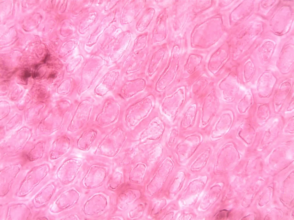
Verocay bodies are the most distinctive feature of Antoni A tissue. They consist of elongated nuclei arranged in two parallel rows separated by seemingly clear zones, according to the Histology Blog review. At low power they read as repeating, orderly bands of nuclei, which is why the term nuclear palisading is used.
Their absence does not exclude schwannoma. The cellular variant predominantly or exclusively shows Antoni A areas and often lacks Verocay bodies, so a pure Antoni A tumor without palisading should still prompt a schwannoma work-up rather than dismissal.
Vessels and stroma
Vascular change is the third structural clue. The Antoni B regions contain ectatic, deformed vessels with hyalinized walls and thrombi, as described by WebPathology. Thick, hyalinized vessel walls in a tumor with loose myxoid areas strengthen the case for schwannoma when Verocay bodies are not obvious.

S-100 and SOX10 staining
Immunohistochemistry confirms what the architecture suggests. Schwann cells consistently show positivity for S-100 and SOX10 (WebPathology). In practice, diffuse staining with both markers supports a Schwann cell lineage, and a panel that includes both is more reassuring than either alone.
Schwannoma versus neurofibroma on histology
The most reliable single discriminator in the supplied teaching material is S-100. The Histology Blog review states that the stain would be intensely positive in a schwannoma and negative in a neurofibroma. Architecture adds support: the paired Antoni pattern, encapsulation and hyalinized vessels point toward schwannoma.
The table gathers the features that guide the decision.
| Feature | Schwannoma finding | Diagnostic use |
|---|---|---|
| Overall behaviour | Slow-growing, encapsulated, benign | Expect a discrete lesion rather than diffuse infiltration |
| Antoni A areas | Cellular, compact | Look here for nuclear alignment |
| Antoni B areas | Hypocellular, spindle cells in loose myxoid or hyalinized stroma | Confirms the biphasic pattern |
| Verocay bodies | Two parallel nuclear rows with clear zones; often absent in cellular schwannoma | Supportive when present, not required |
| Vessels | Ectatic, hyalinized walls, thrombi | Helpful when palisading is scarce |
| S-100 and SOX10 | Consistently positive; S-100 intensely positive versus negative in neurofibroma | Separates schwannoma from neurofibroma |
| Cellular variant mitoses | Usually fewer than 4 per 10 HPF | Low counts fit a benign cellular schwannoma |
Figures and descriptions in the table come from WebPathology and the Histology Blog teaching review.
Vestibular schwannoma pathology
Vestibular schwannoma is the best-known intracranial form. Schwannomas sometimes develop in the eighth cranial nerve, the vestibulocochlear nerve, according to Weill Cornell Neurosurgery. Histologically it is the same tumor as elsewhere, so the Antoni pattern, vessel change and S-100 and SOX10 positivity all apply.
The clinical side differs from the bench. Weill Cornell notes that a schwannoma often requires surgical removal even if it is benign, and that a very small percentage are malignant tumors called neurofibrosarcomas. For the pathologist, that makes confirming benign architecture and low proliferative activity part of every report.
NF2, schwannomatosis and genetic context
Most schwannomas are sporadic. WebPathology gives 90% of cases as sporadic. Among the remainder, Neurofibromatosis 2 accounts for 3%, schwannomatosis for 2%, and multiple meningiomas with or without NF2 for 5%.
That distribution has a practical consequence. A single schwannoma is usually an isolated finding, whereas multiple tumors or a vestibular tumor in a young patient should prompt consideration of NF2 or schwannomatosis. Histology alone does not distinguish the syndromic setting from sporadic disease.
Drug treatment remains an open question. The Congress of Neurological Surgeons guideline discusses targeted therapy for both sporadic and NF2-related vestibular schwannomas. In an everolimus study of 10 NF2 subjects, no reduction in vestibular schwannoma volume was noted over 12 months of therapy.
Related tumor pathology
Nerve sheath tumors are recognised by architecture plus a lineage marker. Other CNS tumors rely on different cues, as in the pages on medulloblastoma histology and craniopharyngioma histology.
Frequently asked questions
What are Antoni A and Antoni B areas?
Antoni A areas are cellular zones; Antoni B areas are hypocellular zones of spindle cells in loose myxoid or hyalinized stroma. They alternate within a schwannoma.
What are Verocay bodies?
They are elongated nuclei arranged in two parallel rows separated by clear zones, found in Antoni A tissue. Cellular schwannoma often lacks them.
How can schwannoma be told from neurofibroma on histology?
S-100 is the clearest discriminator: intensely positive in schwannoma and negative in neurofibroma in the teaching source cited above. Antoni areas and hyalinized vessels favour schwannoma.
What is the role of NF2 in schwannoma?
NF2 is a minority association: 3% of schwannomas in the figures cited above. Most cases are sporadic, and an everolimus study in NF2 patients showed no tumor volume reduction over 12 months.

Leave a Reply