
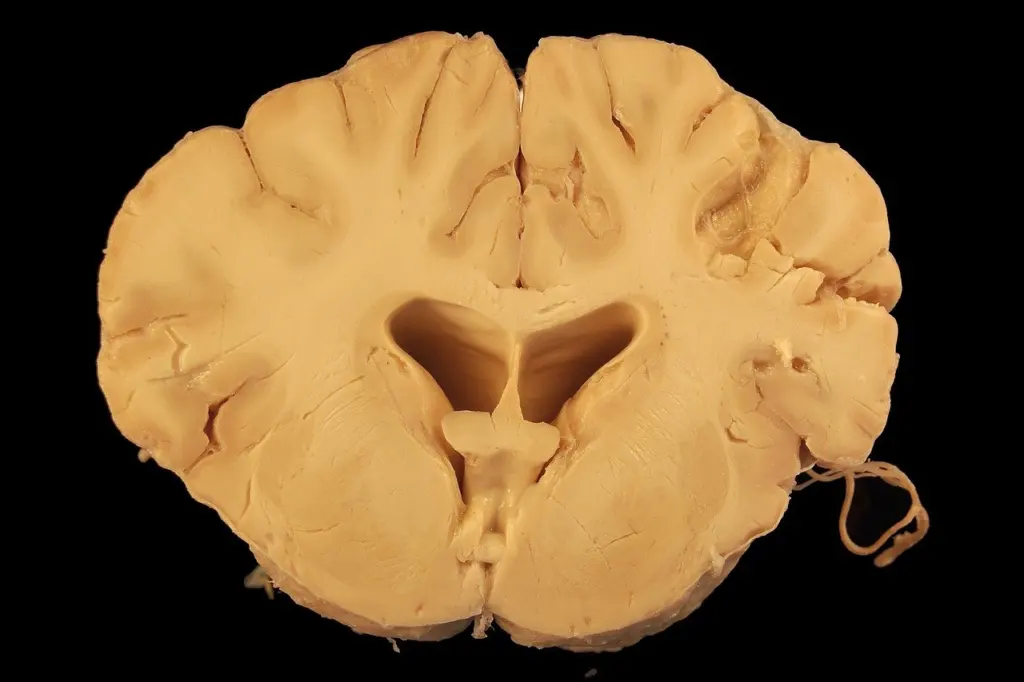
A slide shows dense pink, corkscrew-shaped deposits in glial processes. In astrocytes in neuropathology, that pattern means Rosenthal fibers, and abundant subpial and perivascular deposits point toward Alexander disease, a GFAP-mutation disorder.
This page reads astrocyte pathology from the slide outward, as part of the neuropathology course. It corrects the misunderstandings that most often send students to the wrong diagnosis, then closes with a slide-to-diagnosis checklist.
Astrocytes are not passive filler
Astrocytes are the most abundant glial cell type in the mammalian brain and, according to a peer-reviewed review of astroglial biology, make up up to 50% of brain volume. They supply energy metabolites to neurons and help regulate blood flow and the blood-brain barrier. Protoplasmic astrocytes, the best-characterized type, sit in the gray matter parenchyma. Neurons and glia are compared side by side in neurocytology. Here the question is what changes when astrocytes are injured or carry a defect.
The protein that makes astrocytes easy to find is GFAP, the major intermediate filament protein of astrocytes in the CNS. That is why a GFAP immunostain outlines their stellate processes, and why a GFAP-related disease leaves such a visible mark on astrocytes. The table sets the most common misreadings against what the evidence supports.
| Common misunderstanding | What the evidence supports | Slide-reading use |
|---|---|---|
| Astrocytes are just supporting filler | They supply energy metabolites to neurons and regulate blood flow and the blood-brain barrier, and they make up up to 50% of brain volume. | Treat astrocyte change as a change in tissue function, not as background. |
| Gliosis is one stereotyped reaction | Astrocytes do not react in a stereotyped fashion to all forms of injury and disease. The reaction varies with injury severity and site context. | Describe the pattern and its setting before assigning a cause. |
| GFAP is only a staining marker | It is the major astrocytic intermediate filament protein, and in Alexander disease it accumulates within astrocytes to excessive levels. | GFAP can be both the stain and the disease protein. |
| Astrocytes only react to damage elsewhere | In Alexander disease, dominant mutations in the GFAP coding region start the process within astrocytes themselves. | Consider a primary astrocytopathy when the changes are extensive and the clinical context fits. |
The first two rows draw on a review of astroglial function and injury response, and the last two on a review of GFAP biology in Alexander disease.
Gliosis is not one stereotyped response
Students often treat gliosis as a single, uniform scar. The evidence says otherwise: the astrocytic reaction depends on how severe the injury is and on the context of the site where it occurs. Reactive astrocytes with stellate, GFAP-positive processes tell you that something has provoked the tissue. They do not tell you what.
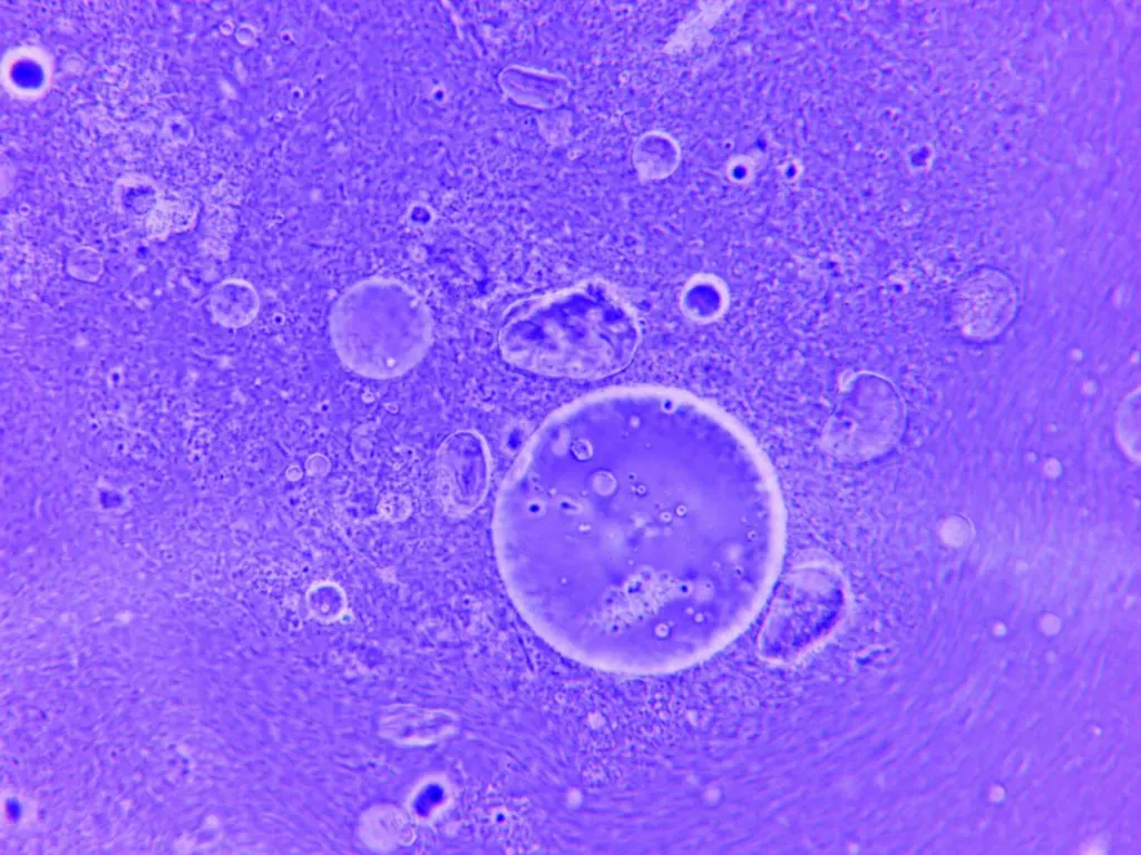
The practical consequence is about wording. A slide report that says only “gliosis” leaves out the information a clinician needs, namely where it is, how it is distributed and what accompanies it. Pair the astrocyte finding with the neighbouring tissue: neurons, vessels, and the other glial responders. Microglia are the other half of that picture, covered in microglia in neuropathology.
Rosenthal fibers: the hallmark inclusion
Appearance on H&E
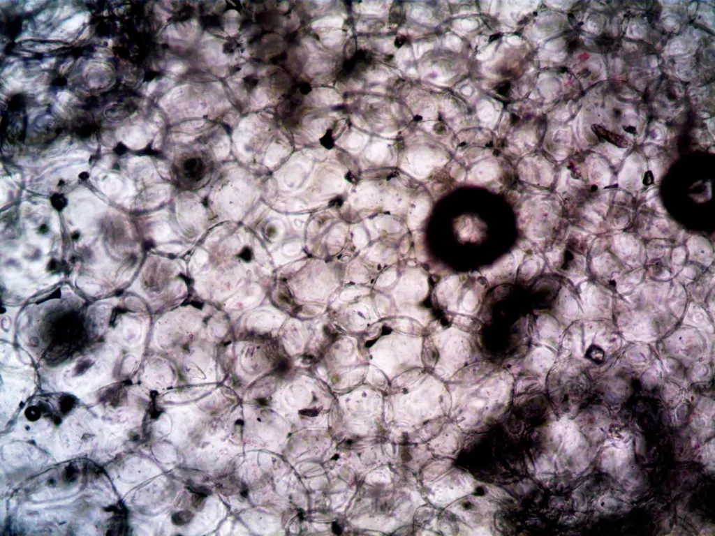
Rosenthal fibers are dense, bright eosinophilic, corkscrew-shaped deposits within astrocyte processes. Their strong pink staining on H&E makes them one of the easier astrocyte inclusions to recognise at low power. Because they sit in processes, they look like elongated or irregular bodies lying among the glial fibres rather than round bodies in the neuropil.
Conditions where they appear
The condition the evidence ties most directly to Rosenthal fibers is Alexander disease. Work catalogued by the Waisman Center Alexander disease research group shows that Alexander disease-associated GFAP mutations induce Rosenthal fiber formation in mice, which links the inclusion directly to the mutant protein.
That link supports a careful rule: the inclusion is a strong prompt, not a verdict. Before naming Alexander disease, check the distribution of the deposits, the clinical picture and, where available, the GFAP genetics. Dense accumulation beneath the pia and around vessels fits the disease histology far better than a few isolated fibers.
Alexander disease: gene, protein and histology
A review in the primary literature describes Alexander disease as a rare, progressive and generally fatal neurological disorder caused by dominant mutations in the coding region of GFAP. A key step in pathogenesis appears to be accumulation of GFAP within astrocytes to excessive levels. In mouse models, the severity of the phenotype correlates with the level of GFAP expression. A 2001 description first linked GFAP mutations to the disease, according to the Waisman group’s publication list.
| Feature | Finding | Slide-reading use |
|---|---|---|
| Gene | GFAP, with dominant mutations in the coding region | Confirms the molecular diagnosis that the histology only suggests. |
| Protein | Glial fibrillary acidic protein, the major astrocytic intermediate filament protein | Explains why astrocytes carry the pathology. |
| Mechanism | Accumulation of GFAP within astrocytes to excessive levels | Links the mutation to the inclusions seen on the slide. |
| Histology | Rosenthal fibers in astrocyte processes, prominent beneath the pia and around vessels | Look at subpial and perivascular zones first. |
| Course | Rare, progressive, generally fatal | Frames the clinical urgency of an accurate diagnosis. |
The gene, protein and mechanism rows come from the GFAP review, and the Rosenthal fiber and mutation link from the Waisman group’s literature listing.
The mechanism explains the histology in plain terms: a mutant filament protein piles up inside astrocytes, and the pile-up shows as eosinophilic inclusions in their processes. The diagnostic chain therefore runs from inclusion, to excess GFAP, to the gene.
Look-alikes: corpora amylacea and Alzheimer type II astrocytes
Corpora amylacea
Corpora amylacea are round, basophilic, laminated bodies, typically seen in subpial or periventricular regions. They are a common age-related finding, so their presence alone should not push a case toward disease. Their blue-purple colour and layered, rounded outline separate them from the pink, corkscrew-shaped Rosenthal fibers.
Alzheimer type II astrocytes
Alzheimer type II astrocytes are recognised by enlarged, pale, vesicular nuclei in gray matter. The change is associated with hyperammonemia, so the question it raises is metabolic rather than structural. Here the abnormality is in the nucleus and the cell looks swollen and pale, whereas in Alexander disease the abnormality is an inclusion in the processes.
From slide finding to likely diagnosis
- Pink, corkscrew-shaped deposits in processes. Think Rosenthal fibers, then ask whether they are abundant beneath the pia and around vessels. If so, Alexander disease and GFAP testing are the next steps.
- Round, basophilic, laminated bodies near the pia or ventricles. Think corpora amylacea. Treat them as a common age-related finding unless the clinical context says otherwise.
- Enlarged, pale, vesicular nuclei in gray matter. Think Alzheimer type II astrocytes and ask about hyperammonemia.
- Stellate, GFAP-positive reactive astrocytes without a distinctive inclusion. Think gliosis. Describe its severity and site, because the reaction is not stereotyped and the cause lies in the surrounding context.
Frequently asked questions
| Question | Answer |
|---|---|
| Is GFAP raised in body fluids in Alexander disease? | According to the GFAP review, levels are consistently elevated in the CSF of patients with Alexander disease, but only occasionally and modestly elevated in blood. |
| Has any treatment been tested for Alexander disease? | The Waisman group’s literature listing includes a 2021 study in which antisense therapy reversed GFAP pathology in a rat model. That is animal research, not an established treatment for patients. |
| How do I tell corpora amylacea from Rosenthal fibers? | Use colour and shape. Corpora amylacea are basophilic, round and laminated, while Rosenthal fibers are bright eosinophilic and corkscrew-shaped within astrocyte processes. |
| What does the term Alzheimer type II astrocyte describe? | It describes a cell with an enlarged, pale, vesicular nucleus in gray matter. The change is associated with hyperammonemia. |

Leave a Reply