
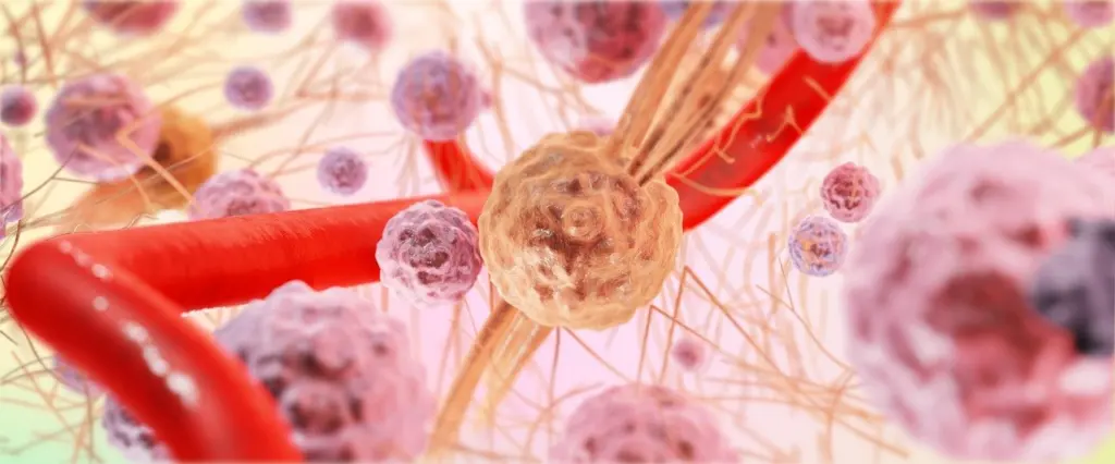
Quick answer: Oligodendroglioma histology shows round nuclei, perinuclear halos and chicken wire capillaries, but these look-alike features only support the diagnosis. IDH mutation plus 1p/19q codeletion confirms it.
Slide appearance, molecular results and grade each answer a different question, so read them in that order. This page sits within our wider brain tumor pathology coverage. For contrast with other glioma and ependymal patterns, see pilocytic astrocytoma histology and ependymoma histology.
What confirms the diagnosis
Oligodendroglioma, IDH-mutant and 1p/19q-codeleted, is defined by its genetics as well as its microscopy. The Brain Tumour Charity states that two genetic alterations confirm the diagnosis: an IDH1 or IDH2 mutation and 1p/19q co-deletion. The American Brain Tumor Association describes the same pairing: mutations in IDH1 or IDH2 and loss of the chromosomal arms 1p and 19q.
Use this order of trust when working a case:
- Start with the slide. Round nuclei, halos and delicate capillaries tell you what to test for. They do not settle the diagnosis alone.
- Then test the molecular pair. The tumour needs both an IDH mutation and 1p/19q codeletion. One without the other points elsewhere.
- Grade last. Once the tumour is established as an oligodendroglioma, grade 2 versus grade 3 is decided by slide features such as mitoses, cellularity, microvascular proliferation and necrosis.
Oligodendroglioma histology under the microscope
Oligodendrogliomas arise from oligodendrocytes, a glial cell type, according to the American Brain Tumor Association. The tumour cells are described as having short arms or a fried-egg shape. The OncoDaily review lists the key microscopic features as round nuclei, a perinuclear halo and a branching capillary network.
Round nuclei and perinuclear halos
A perinuclear halo is a clear zone around a round nucleus. Together they produce the fried-egg look. This is the feature most often associated with the tumour. Treat it as a cue to order molecular testing rather than as proof, for the reasons covered in the pitfalls section below.
Chicken wire vasculature
Chicken wire vasculature is a pattern of delicate, branching capillaries running through the tumour. OncoDaily gives it as the vascular signature of oligodendroglioma, written as a “chicken wire” pattern. On a slide, look for a fine, interlacing network of thin vessels between the tumour cells. This pattern is distinct from the proliferating vessels that mark higher grade, covered in the grading section.

Calcification and other tissue findings
The American Brain Tumor Association notes that these tumours often contain calcifications, areas of haemorrhage and cysts. OncoDaily adds that calcifications are better seen on CT scans. Finding calcification in tissue is therefore consistent with the diagnosis and with what imaging often shows, but it is a supporting finding only.
Molecular confirmation
Molecular results turn a suggestive slide into a diagnosis. The two required alterations are tested with different methods.
1p/19q codeletion
1p/19q codeletion means loss of both the 1p and 19q chromosomal arms. It is required because it separates oligodendroglioma from other IDH-mutant gliomas. OncoDaily reports that under the WHO 2021 classification, IDH-mutant tumours that lack the codeletion are classified as astrocytomas. The Brain Tumour Charity says that if the codeletion is absent the tumour may be another glioma type. OncoDaily lists fluorescence in situ hybridization (FISH), PCR and next-generation sequencing as ways to confirm it.
IDH1 mutation
IDH1 is the more commonly named of the two IDH genes, with IDH2 as the alternative. OncoDaily notes that IDH mutation is usually tested by immunohistochemistry or sequencing. An immunostain gives a quick, slide-based readout, while sequencing covers the wider set of possible mutations.
TERT promoter mutation
The Brain Tumour Charity states that a TERT promoter mutation found together with 1p/19q co-deletion and an IDH-mutant result suggests oligodendroglioma. It adds weight to the pair but does not replace it.
Grade 2 versus grade 3
Grade 2 and grade 3 oligodendrogliomas differ in how aggressive they look on the slide and how they behave. OncoDaily describes grade 2 as having fewer mitoses and no microvascular proliferation or necrosis. Grade 3 shows increased cellularity and mitotic activity, and sometimes microvascular proliferation and necrosis.

| Feature | Grade 2 | Grade 3 | Planning use |
|---|---|---|---|
| Cellularity and mitoses | Fewer mitoses | Increased cellularity and mitotic activity | Count mitoses first; this is the usual starting point for separating the grades |
| Microvascular proliferation and necrosis | Absent | Sometimes present | Their presence points to grade 3, but their absence does not rule it out |
| Growth behaviour | Slower growth | Grows faster and is more likely to spread within the brain | Explains why the grade changes how closely a tumour is managed |
Histologic features are from OncoDaily, and growth behaviour is from the Brain Tumour Charity.
The American Brain Tumor Association also associates grade 3 with more rapid growth. Those genetic findings are supporting observations. The grade itself rests on the slide features in the table.
Mitoses and cellularity can vary across a tumour, so a grade assigned from a small sample may understate the more active areas. Grade after reviewing as much tissue as is available.
Typical age and location
Sources give overlapping but not identical age windows. The American Brain Tumor Association says most cases occur between the ages of 20-40. The Brain Tumour Charity points to ages 30-50. Medbullets gives 25-45. Read these as a young to middle adult range rather than a fixed cutoff. Both the American Brain Tumor Association and the Brain Tumour Charity describe the tumour as slightly more common in men.
The frontal and temporal lobes are common locations. The Brain Tumour Charity adds that the majority occur in the frontal lobe, followed by the temporal and then the parietal lobe. A frontal or temporal mass in a younger adult fits the pattern, but it should not substitute for the slide and molecular work.
Pitfalls: when look-alike features mislead
Perinuclear halos can mislead if they are treated as diagnostic. The diagnosis depends on the molecular pair, so a tumour with a fried-egg appearance and no 1p/19q codeletion may be another type of glioma. When IDH is mutated but the codeletion is absent, OncoDaily reports the tumour is classified as an astrocytoma under WHO 2021.
- Classic look, no codeletion. Do not call it oligodendroglioma. Review the molecular result and consider an IDH-mutant astrocytoma.
- Looks circumscribed on imaging. Do not assume the tumour is confined. It is diffuse in the brain tissue, as described by the Brain Tumour Charity.
- Grade 2 features on a small sample. Be cautious about excluding grade 3 when little tissue is available, since the high-grade features can be focal.
Frequently asked questions
How common is oligodendroglioma?
The Brain Tumour Charity says oligodendrogliomas account for roughly 3% of all primary brain tumours.
Why can an oligodendroglioma look well defined on a scan?
OncoDaily notes that these tumours are often well circumscribed on imaging but histologically diffuse, so the visible edge does not show how far tumour cells extend.
Does an IDH mutation affect treatment response?
The Brain Tumour Charity states that IDH-mutant tumours have a better response to treatment.
Which cells does oligodendroglioma come from?
It arises from oligodendrocytes, a type of glial cell, according to the American Brain Tumor Association.
Leave a Reply