
Quick answer: Peripheral nerve pathology is easiest to learn by asking whether the axon or the myelin is the primary target. Axonal injury ends in Wallerian degeneration. Demyelination strips myelin from an axon that stays intact.
Start with that split before memorising named diseases. The same logic carries through the wider neuropathology course, and the rest of this guide applies it to injury, acquired neuropathy and inherited neuropathy.
Choosing the pattern: axon first or myelin first
Peripheral nerve injury is common enough to matter in everyday practice. Orthobullets gives an incidence of 16.9 per 100,000 per year. Whatever the cause, a nerve can react in only a few ways, and the first decision is where the damage sits.
- Myelin is the target. Schwann cells make the myelin that insulates peripheral axons and raises conduction velocity, so losing it slows or blocks conduction. In this pattern the axon remains intact.
- The axon is the target. The axon and its myelin are both disrupted, and the segment beyond the injury degenerates. Recovery depends on regrowth and is hard to predict.
- The whole nerve is divided. Every layer is disrupted, and no recovery should be expected without surgical repair.
- Compression sits between these. Pressure can block conduction without cutting anything. Chronic compression also drives Schwann cell proliferation and apoptosis, so the picture changes with duration.
Myelin disruption typically occurs before axon disruption. A nerve that begins as a demyelinating problem can therefore progress to axonal loss if the insult continues.
Injury patterns compared
The Seddon classification is the most useful frame for matching histology to prognosis. The table sets each grade beside what you would expect to see and what it means for a pathology or electrodiagnostic read.
| Pattern (Seddon term) | Histology | Electrodiagnostic finding | Outlook | Planning use |
|---|---|---|---|---|
| Segmental demyelination (neurapraxia) | Focal, temporary demyelination with the axon intact; no Wallerian degeneration | Conduction velocity slowing or block, without fibrillation potentials | Reversible | Look for a focal myelin lesion and expect recovery if the cause is removed. |
| Axonal disruption (axonotmesis) | Axon and myelin sheath disrupted, with focal conduction block and Wallerian degeneration | Fibrillations and positive sharp waves on EMG | Recovery unpredictable | Treat as true axon loss. Counsel cautiously and follow regrowth over time. |
| Complete division (neurotmesis, Sunderland grade V) | Nerve completely severed, with all layers disrupted | Axonal pattern | No recovery unless surgical repair is performed | Plan for surgical repair rather than waiting for spontaneous return. |
These grades and findings follow the nerve injury summary on Orthobullets.
Axonal injury and Wallerian degeneration
After a transection, the distal segment undergoes Wallerian degeneration, in which phagocytes degrade the axoplasm and myelin. This is the defining event of axonal injury and is absent in simple demyelinating conduction block.
Regrowth sets the timescale. Proximal budding occurs after about 1 month and produces sprouting axons that migrate at 1 mm/day, according to Orthobullets. Treat that as a typical regrowth speed, not a guaranteed one. It is the reason a long nerve takes far longer to recover than a short one.
Compression shows how pattern and dose interact. Pressure above 60 mm Hg can completely block conduction, while lower pressure such as 30 mm Hg causes paraesthesias. The same source notes that chronic compression produces Schwann cell proliferation and apoptosis, so the histology of a long-standing entrapment differs from that of a brief insult.
Some injuries are predictable. Correction of a valgus deformity in total knee arthroplasty can lead to common peroneal nerve palsy.
Segmental demyelination and onion bulb formation
Segmental demyelination is loss of myelin along individual internodes while the axon is preserved. In teased fibre preparations it appears as bare axon segments between stretches of normal myelin. Because Schwann cells make the myelin that speeds conduction, the clinical and electrical effect is slowing or block rather than the fibrillation seen in axon loss.
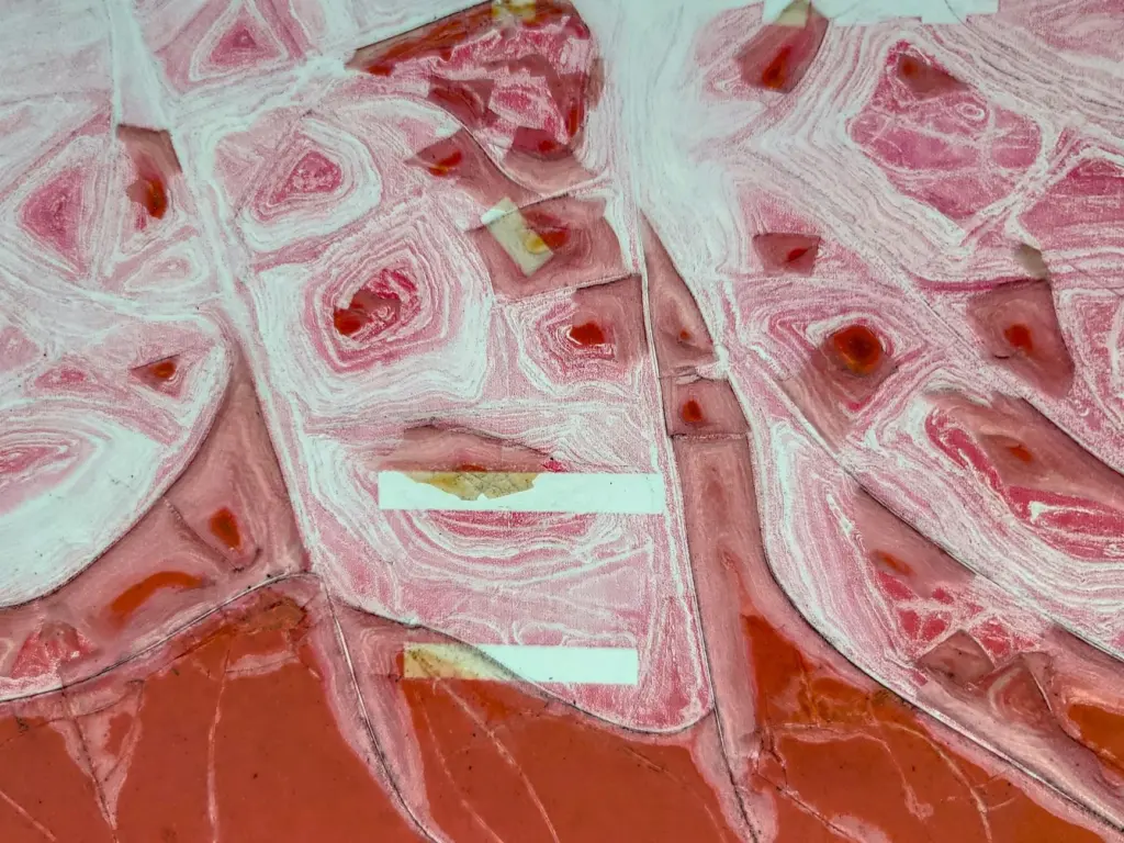
Onion bulbs are the long-term record of Schwann cell activity. They are concentric layers of Schwann cell processes wrapped around a nerve fibre in cross-section. This fits the finding that chronic compression leads to Schwann cell proliferation, so onion bulbs suggest a long-standing process rather than a single acute insult. A section with many of them points you toward a chronic demyelinating or inherited neuropathy.

Demyelination is not unique to the peripheral nervous system. For the central counterpart, where the myelin-forming cell is different, see progressive multifocal leukoencephalopathy pathology.
Acquired example: Guillain-Barré syndrome
Guillain-Barré syndrome pathology is the acquired example usually used to teach inflammatory demyelination. The nerve shows endoneurial inflammation, with macrophages stripping myelin from fibres. It fits the demyelinating pattern in the table: myelin is attacked first, and electrical slowing or block follows.
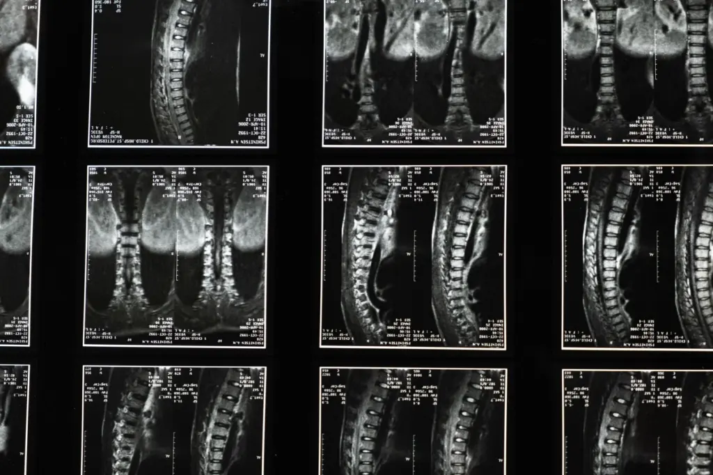
When reading a section, look for inflammatory cells within the endoneurium and for myelin breakdown products. A purely axonal picture without that inflammatory demyelination argues for a different cause.
Inherited example: Charcot-Marie-Tooth and PMP22
Charcot-Marie-Tooth pathology teaches the same patterns from the genetic side, and PMP22 is the gene to know first. Dosage changes in PMP22 produce opposite diseases. According to Charcot-Marie-Tooth News, hereditary neuropathy with liability to pressure palsies (HNPP) is caused by PMP22 deletion, the opposite of the PMP22 duplication that causes CMT1A. The same report states that missense mutations in PMP22 have been linked to different types of peripheral neuropathy.
- CMT1A. It is caused by PMP22 duplication, so the nerve carries an extra copy of the gene.
- HNPP. It is caused by PMP22 deletion. The name fits the compression findings above, because pressure palsies are the characteristic problem.
- PMP22 missense variants. These sit on the same spectrum and can produce different neuropathy types.
For your own revision, start with gene dosage (duplication versus deletion) and only then add individual variants. Both directions of PMP22 dosage affect Schwann cell myelin, which is why these disorders belong with the demyelinating pattern.
Linking morphology to the muscle and the clinic
Nerve disease is often first recognised through its effect on muscle. After axonal loss, denervated fibres atrophy in groups, and reinnervation produces fibre type grouping. The muscle therefore records how long and how completely the nerve has been affected.
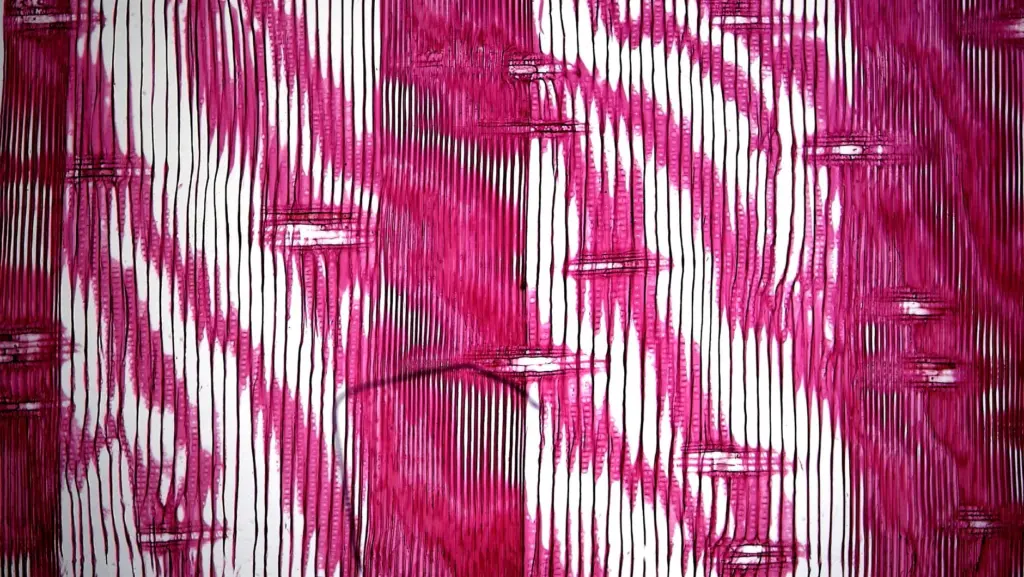
Electrodiagnosis ties the morphology to the patient. A pattern of conduction velocity slowing or block without fibrillation potentials points to demyelination. Fibrillations and positive sharp waves on EMG point to axonal forms. Neither result replaces histology, but together they tell you which pattern to look for before the biopsy is cut.
Troubleshooting a pattern call
- Slowing but weakness persists. Because myelin disruption typically precedes axon disruption, an initially demyelinating nerve may be progressing to axonal loss. Re-check for EMG evidence of fibrillation.
- Many onion bulbs, little inflammation. This favours a chronic or inherited process over an acute inflammatory one. Consider the PMP22 dosage disorders and compression history.
- Axonal picture, no clear trauma. Axon loss with Wallerian degeneration can follow a focal lesion upstream, so look for a compressive or mechanical site.
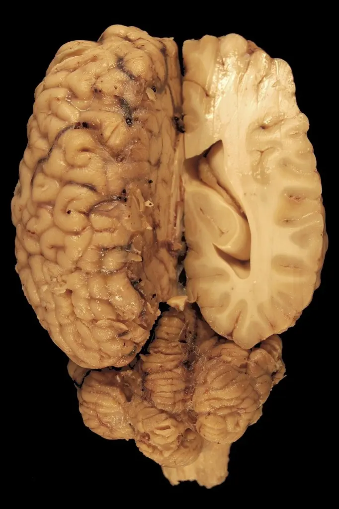
- Handling tissue. Nerve and muscle sampling is a separate workflow from the brain. For the central tissue side, see neuropathology brain dissection.
Frequently asked questions
Is PMP22 linked to painful neuropathy?
Yes, in at least one report. A PMP22 T118M variant was described in three unrelated patients with painful neuropathy, with CMT scores from 2/28 to 8/28. The first patient was a 64-year-old man with a burning sensation in his feet.
Does demyelination always spare the axon?
No. In neurapraxia the axon stays intact and recovery is reversible. But myelin disruption typically comes before axon disruption, so continued injury can damage the axon as well.
Which nerve is at predictable risk during knee surgery?
The common peroneal nerve. Realigning a valgus knee during total knee arthroplasty is a recognized setting in which the common peroneal nerve may be injured, producing palsy.
Leave a Reply