

A patient on natalizumab develops weakness in one arm, and MRI shows white matter lesions. Progressive multifocal leukoencephalopathy pathology explains the link: JC virus infects and kills oligodendrocytes and astrocytes, and myelin is progressively destroyed.
This case study follows one viral demyelinating disease from the infected cell to the MRI lesion to the tissue section. It is one chapter of the wider neuropathology course, which sets PML beside other central nervous system diseases.
From JC virus exposure to infected glial cells
PML is a serious viral infection of the brain, and its cause is a virus most adults have already met. The same fact sheet reports that between 80–85% of adults are exposed to JC virus worldwide. An encyclopedia reference on PML puts exposure among US adults at at least 80%. Exposure is therefore the baseline state, and disease is the exception.
What separates disease from exposure is reactivation. Neurology review commentary describes PML as caused by reactivation of the polyomavirus JC. In immunocompromised people, the encyclopedia reference says, the reactivated virus travels through the circulatory system to the brain. After prolonged immune compromise it changes into a form that can reach brain tissue and cause disease.
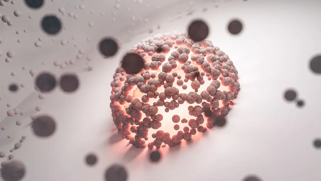
Once in the brain, JC virus infects and kills two kinds of glial cell, oligodendrocytes and astrocytes. Infection of these cells is followed by progressive destruction of myelin, the substance that wraps around nerve fibers. The lesion described in the rest of this article is the visible result of that chain: reactivation, spread through the blood, glial infection, then myelin loss.
Common misunderstandings about PML
Several assumptions about PML are tempting and wrong. The table sets each against what the evidence supports and what that means in practice.
| Misunderstanding | What is supported | Practical meaning |
|---|---|---|
| PML is a new infection caught by weakened patients | 80–85% of adults worldwide are exposed to JC virus; disease follows reactivation | Past exposure does not predict PML. Immune status is the deciding condition. |
| PML is only an HIV complication | In an older reference figure, most cases occur with lymphatic cancers, and HIV is one of several settings | Think of PML in any person with significant immunosuppression, not one diagnosis. |
| Only myelin is damaged | The virus infects and kills oligodendrocytes and astrocytes; myelin is progressively destroyed | The lesion reflects loss of two glial cell types plus myelin. |
| An abnormal MRI settles the diagnosis | Symptoms plus white matter destruction on CT or MRI support it; CSF PCR may show JC virus DNA; only brain biopsy is absolutely definitive | Imaging raises the question. CSF testing or tissue answers it. |
These contrasts draw on the IAPAC fact sheet on PML and the encyclopedia reference cited above.
The first misunderstanding is the most consequential. Because exposure is so common, a positive history of JC virus contact is not a reason for alarm in itself. It matters only when immune defenses are compromised enough for the virus to reactivate.
Tissue changes and demyelination
Under the microscope, PML is a disease of white matter in which infected glia die and myelin disappears. Because the virus infects oligodendrocytes and astrocytes, the tissue picture involves both cell populations, not myelin alone. The destruction is progressive, which is why the lesion is a record of ongoing infection rather than a single injury.
Special stains make the myelin loss visible. A Luxol fast blue section stains intact myelin, so patchy pale areas at the gray-white junction mark where myelin has gone. Reading that pattern next to the cellular findings is how a pathologist links the lost myelin to the infected cells.

Tissue is also the final arbiter. The encyclopedia reference states that only brain biopsy can result in an absolutely definitive diagnosis. For a disease whose clinical and imaging features can overlap with other white matter conditions, that standard explains why biopsy remains the reference point.
MRI compared with tissue findings
MRI shows where white matter has been destroyed, and tissue shows why. The clinical diagnosis rests on symptoms plus evidence of white matter destruction visualized on CT or MRI. The IAPAC fact sheet likewise notes that PML can be diagnosed using a scan of the brain by MRI.
The scan and the section describe the same lesion at different scales. On FLAIR imaging the lesions appear as asymmetric subcortical white matter abnormalities without mass effect. At the microscope, the same territory shows lost myelin and infected glia. A lesion that looks like a space-occupying mass would suggest a different process, so the absence of mass effect is a useful reading cue.

The limit of MRI is that it maps damage without identifying the cause. It does not by itself show JC virus. That is why a suggestive scan in an immunosuppressed patient is followed by virologic or tissue confirmation, covered next.
Confirming the diagnosis by CSF testing or biopsy
Confirmation uses either spinal fluid or brain tissue. Cerebrospinal fluid can be tested with polymerase chain reaction, and these specialized tests may demonstrate the presence of JC virus DNA. The IAPAC fact sheet confirms that the diagnosis can be made by MRI or by testing of the spinal fluid.
The choice between the two routes follows a simple logic:
- CSF PCR first. It suits a patient whose symptoms and white matter lesions fit PML, because it looks directly for viral DNA without sampling brain.
- Biopsy for certainty. It suits the situation where non-invasive results are not enough, because tissue is the only route to an absolutely definitive diagnosis.
In tissue, an immunostain for SV40 large T antigen marks infected oligodendrocyte nuclei, which shows the virus itself in the cells that carry it.
Which patients are at risk
PML risk follows immunosuppression, whatever its source. The settings below are the ones the evidence ties to the disease.
- HIV and AIDS. Researchers estimate that about 6% of people with AIDS develop PML, and most cases appear at CD4 counts below 100 cells/mm3. An older reference estimate gives 2–7% of people with HIV, a broader group with a wider range.
- Monoclonal antibody drugs. Neurology commentary links natalizumab, efalizumab and rituximab to PML. Anyone treated with these drugs, and the clinicians following them, should treat new neurologic symptoms as a reason to consider PML.
- Lymphatic cancers. An older reference figure says 62.2% of cases occur in people with lymphatic cancers, making this the largest group in that dataset.
- No other cause. The same reference reports that only 5.6% of cases occur with no other underlying source of immunocompromise. PML without a known immune problem is uncommon, though not impossible.
The older percentages come from a reference text and differ from newer estimates, so treat them as a picture of the pattern rather than current rates. The consistent message is that the immune system, not the virus alone, decides who becomes ill.
Outlook, treatment and related pathology
PML is fatal in about 50% of cases according to the IAPAC fact sheet. An older reference text described it as uniformly fatal, usually within one to four months of the initial symptoms. The difference between those statements reflects how prognosis has changed, so the more recent figure is the better guide.
There is no approved treatment for PML, although several treatments may be helpful. Because the disease depends on failed immune control, the most promising approach is strengthening the immune system, such as with antiretroviral therapy in HIV. That explains why identifying and addressing the underlying immunosuppression matters as much as confirming the diagnosis.
For comparison with a bacterial infection of the meninges, see suppurative meningitis pathology. For myelin disease outside the brain and spinal cord, see peripheral nerve pathology.
Frequently asked questions
| Question | Answer |
|---|---|
| What are the first symptoms of PML? | The IAPAC fact sheet says the first symptoms are weakness or coordination problems in an arm or leg. |
| Do people with HIV and PML survive? | Many do. The same fact sheet reports that more than half of people with HIV and PML now survive for at least two years. |
| Is PML caused by a newly acquired infection? | Usually not. It is caused by reactivation of JC virus in people whose immune defenses are compromised, according to neurology review commentary. |


Leave a Reply