
A neuropathology course works best when you start with the cells and normal histology, then move through injury, infection, tumors and neuromuscular disease. Choose your first block by what your syllabus tests soonest, and keep a slide-based routine running throughout.
The first decision is whether you need normal structure before disease. If cell types and histology feel unfamiliar, begin with neurocytology. If your timetable has already reached injury, infection or tumors, go straight to that block and return to the cell material as gaps appear.
Choosing where to start
Three questions settle the entry point. What does your syllabus assess next? How comfortable are you with normal histology? Do you have access to slides and specimens, or only lecture material? The answers matter more than any fixed order, because the blocks of a neuropathology course lean on each other in predictable ways.

The University of Pittsburgh’s basic neuropathology lecture list is a useful reference for the shape of the subject. It includes neuroanatomy and histology, muscle and nerve pathology, glial and non-glial tumors of the CNS, hypoxia and ischemia, malformations, hydrocephalus, and viral and non-viral infections. Each of those can serve as a starting point for a different kind of student.
- Start with cells if histology is unfamiliar. Neurons and glia underpin nearly every later block. Reading about neurocytology first means that later slides have recognizable landmarks.
- Start with injury if your term is organized by mechanism. Hypoxia, ischemia, pressure and trauma are taught as patterns of damage. Learning one pattern well gives you a template for the next.
- Start with infection if you are learning from cases. Meningitis and other CNS infections are usually presented as clinical scenarios, so the pathology is easier to place if you begin from the presentation.
- Start with tumors only after glia. Pitt divides its tumor lectures into glial and non-glial groups. Knowing the glial cell types makes the first group much easier to organize.
- Start with nerve and muscle if your lab is hands-on. Biopsy-based topics reward early practice with slides, and peripheral nerve pathology is a self-contained place to build that habit.
Wait on a block when it depends on one you have not covered. Tumor classification before glial biology, or perinatal injury before general hypoxic-ischemic damage, usually means relearning the material later. The sections below follow the order that avoids most of that backtracking.
How a neuropathology course is organized from cell biology to disease
A typical neuropathology course moves from normal structure to disease mechanism to named conditions. Pitt presents its basic lectures as a named list with video links, so a student can follow the sequence or jump to a single topic. That format suits revision, since you can return to one lecture without replaying the rest.
The lecture-based sequence
Pitt’s basic list opens with neuroanatomy and histology and includes muscle and nerve pathology, tumors, hypoxia and ischemia, malformations, hydrocephalus and infections. Read as a course map, it falls into a few families: normal structure, developmental and fluid disorders, injury, infection, neoplasia, and nerve and muscle disease. The table later on this page lays those families out side by side.
Patterns in catalogue courses
Course catalogues from other programs show the same spine, even when the content is broader or narrower than a medical-school pathology block. They are not all neuropathology courses, but they show how teachers frame neurological disease.
- Structure, function, then disease. Alvernia University’s graduate Fundamentals of Neurology is a 4-credit course. It covers anatomy, physiology, pathophysiology, epidemiology, etiology and presentation.
- Brain anatomy as the entry to disease. The University of Southern California’s MEDS 340 is a 2-unit course offered in fall and spring terms. It examines neurological diseases by teaching the anatomy and physiology of the human brain.
- Common conditions as case studies. Oklahoma City Community College’s PTA 2024 is a 4-credit-hour course. It uses conditions such as cerebrovascular accidents, traumatic brain injury, multiple sclerosis, Parkinson’s disease and spinal cord injury to teach pathophysiology.
- Problem-based learning. Eastern Michigan University’s PAS 640 is a 3-credit-hour course on infectious disease, ENT and cardiology topics. It integrates anatomic and physiologic principles through problem-based learning, which is a model you can borrow for neuropathology revision.
The common thread is that structure comes first, mechanism second and named disease third. If a resource skips straight to named diseases, supply the structure and mechanism yourself by returning to the cell and histology material.
Cells and normal histology
The first block teaches you to recognize what is normal so that abnormal findings stand out. Neurons and glia are the cells to know, and the questions to ask of each are the same: what does it look like on a routine stain, what does it contribute to normal function, and how does it change when tissue is injured?
Start with the overview in neurocytology, then split into the two glial cell types that recur through the rest of the course.
- Astrocytes. Read astrocytes in neuropathology to see how this cell is described in injury, since many later chapters refer back to its reaction.
- Microglia. Read microglia in neuropathology for the link between this cell type and inflammatory change in the nervous system.
Spend the most time here if you plan to study from virtual or glass slides. A student who can name the normal cells in a cortical section reads an abnormal section faster, because the question shifts from “what is this?” to “what has changed?”. If you are short on time, a brief pass through this block followed by an immediate move to injury is better than a long delay before disease.
Patterns of CNS injury
The main patterns of CNS injury that a course teaches are hypoxic and ischemic damage, vascular disease, raised pressure with herniation, fluid disorders such as hydrocephalus, and trauma. Pitt’s list names hypoxia and ischemia, hydrocephalus and a forensic series that includes blunt force trauma. Think of each pattern as a recurring way the brain responds to a different insult.
Hypoxia and ischemia
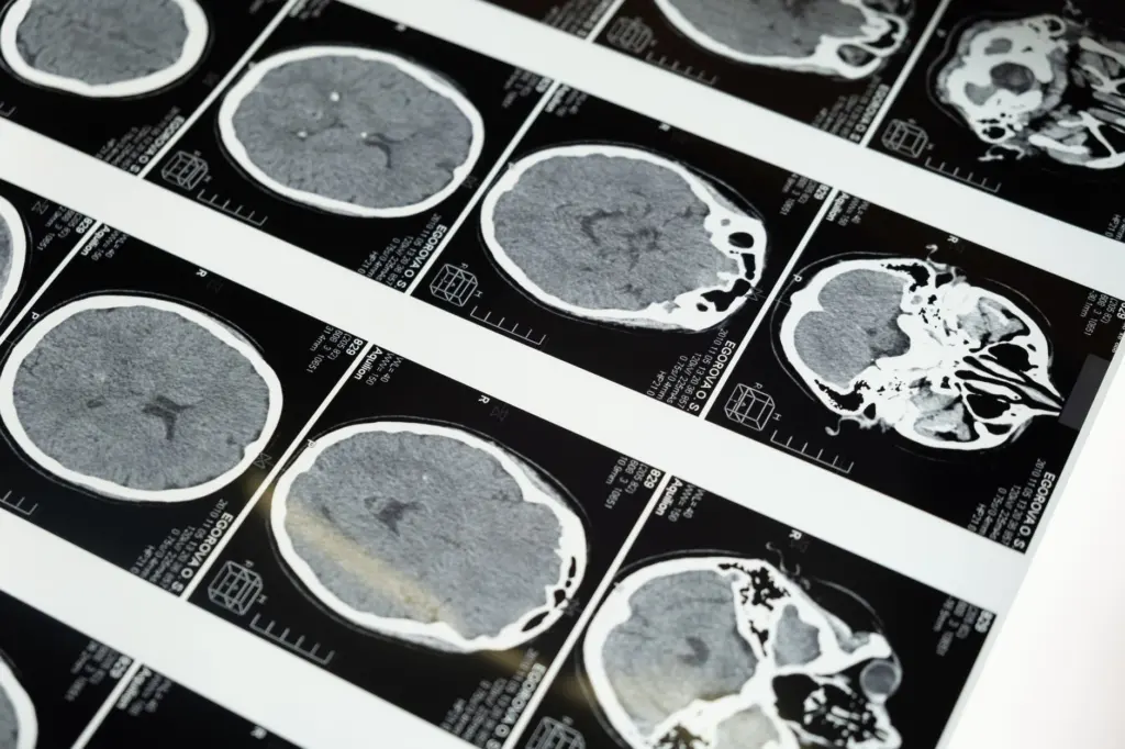
Start with the general framework in hypoxic ischemic encephalopathy neuropathology, because hypoxic-ischemic encephalopathy is the reference example for this pattern. Pitt lists hypoxia and ischemia as its own lecture, which shows how central the topic is to a basic course.
The perinatal material branches from this foundation. Once the general picture is secure, move to perinatal asphyxia brain pathology, then periventricular leukomalacia pathology and kernicterus neuropathology. Taking them in that order keeps the adult and neonatal stories from blurring.
Vascular disease
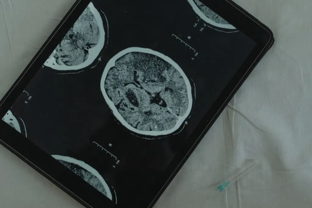
Stroke and related vessel disease overlap with hypoxia and ischemia but are taught as their own topic. Read cerebrovascular disease pathology after the hypoxic-ischemic material, so you can separate what is shared from what belongs to vessel disease. Oklahoma City Community College also includes cerebrovascular accidents among the common conditions it analyzes, which reflects how routinely this subject appears in neurological teaching.
Pressure, herniation and hydrocephalus
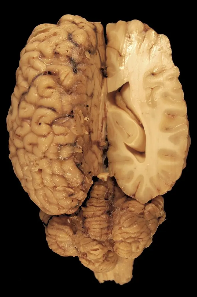
Raised pressure inside a fixed skull is the topic that links several chapters. The overview of brain herniation types is where to learn the gross patterns, and it pairs naturally with your gross specimen sessions, because herniation is visible to the naked eye on a cut brain.
Hydrocephalus is a distinct entity in Pitt’s list. It appears as its own lecture, directly alongside adult and pediatric malformations. Treat it as a bridge between developmental disorders and pressure-related disease: you may meet it in either block, and keeping it in both mental folders helps when an exam question crosses the boundary.
Trauma
Trauma teaching is often split between clinical and forensic perspectives. Pitt offers a forensic neuropathology series that covers blunt force trauma and gunshot wounds, a reminder that the mechanism of injury shapes what you see. Start with traumatic brain injury neuropathology for the core patterns, and use forensic material as an extension if your course includes it.
Seizure-related change
Seizure disorders sit slightly apart from the injury patterns above, because the question is often what structural change underlies the clinical problem. Epilepsy neuropathology is best approached after you can recognize hypoxic and ischemic damage, since those findings recur in its differential.
Infection and inflammation
Infections of the CNS are taught as a distinct block, and Pitt splits it into acute and chronic viral infections and non-viral infections, each with video lectures. That split gives you a practical way to organize the material: ask first whether the agent is viral or non-viral, then ask whether the course is acute or chronic.
Meningitis is the entry case for the non-viral group. The deeper page on suppurative meningitis pathology covers bacterial meningitis alongside brain abscess, and it works best read next to a slide of the meningeal exudate so that the words attach to an image.
For the viral group, progressive multifocal leukoencephalopathy pathology is a good example of a chronic viral process affecting white matter. Read it after the microglia material, because the inflammatory cells explain much of what you see in tissue.
If your course uses problem-based learning, as Eastern Michigan’s catalogue entry describes for its infectious disease course, build a small comparison grid yourself. Put agents down one side and the questions that the case will force you to answer across the top: which structures are involved, what the cellular response looks like and how the course evolves.
Tumors of the CNS
CNS tumors are the block where classification matters most. Pitt lists glial tumors and non-glial tumors of the CNS as separate lectures, and that division is the simplest organizing principle for a beginner: first decide which cell family a tumor belongs to, then learn the individual entities within it.
Learn the glial group only after you are comfortable with astrocytes and the other glial cells, since the tumor names are built on the cells of origin. For the non-glial group, concentrate on how each entity differs from the glial tumors, rather than memorizing a flat list. A short table of features you build yourself, with columns for cell of origin, typical location and microscopic appearance, will serve you better than rereading chapters.
Nerve and muscle disease
The neuromuscular block moves outside the brain. Pitt lists muscle pathology and nerve pathology as separate lectures among its basic topics, so you can expect them to be taught as biopsy-based subjects. The nerve page, peripheral nerve pathology, is the place to learn how a nerve is examined and what its disease patterns look like.
This block is a good place to build slide discipline. Muscle and nerve sections reward a systematic look at the tissue rather than a search for a single obvious lesion. Practice describing what you see before you name a diagnosis. If your program offers lab rotations, this is an excellent block to prioritize them for.
Studying slides and gross specimens effectively
The most effective study method pairs a slide or specimen with a written description in your own words before you check the answer. Pitt offers medical student lab rotations and a virtual microscopy resource covering disease entities, which together support the hands-on and the self-directed sides of study.
Slides
Work from overview to detail, and describe before you diagnose.
- Identify the tissue and the stain, and decide whether the section looks normal or abnormal.
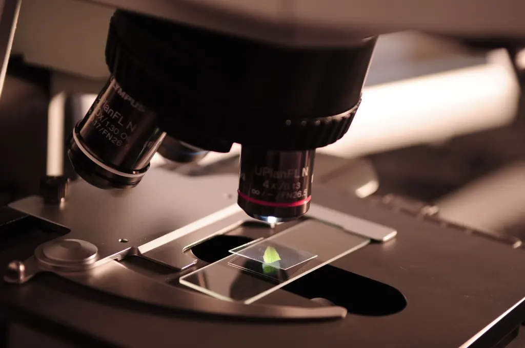
- Scan at low magnification for the distribution of change before moving to high magnification for cell detail.
- Name the cell types you can recognize, using the cell material from the first block.
- Write a one-line description, then compare it with the answer in the lecture or atlas.
- Return to the same entity a few days later and describe it again without notes.
Gross specimens
Gross study works on the same principle: describe, then interpret. Use herniation, infarction and other patterns as visual anchors, and connect each to the mechanism you read about. For a deeper look at how brains are examined, neuropathology brain dissection explains the approach to cutting and sampling that produces the specimens you are shown.

Studying in groups
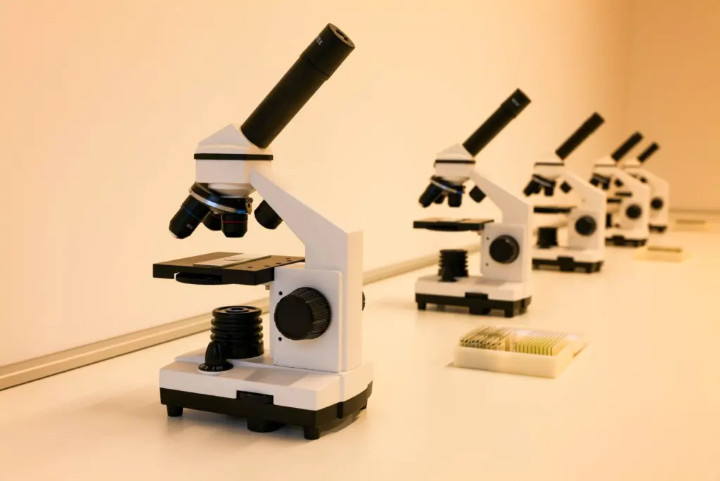
Group sessions help because describing a slide aloud exposes gaps that silent reading hides. Take turns being the describer and the checker, and rotate the role with each new slide. This works with a shared screen of virtual slides just as well as with a multi-headed microscope.
Course map at a glance
The table below turns the lecture list into a planning tool. Use it to decide which block to open next and what to pair it with.
| Course block | Topics in the lecture list | Planning use |
|---|---|---|
| Normal structure | Neuroanatomy and histology | Open first if cell types are unfamiliar; return to it whenever slides confuse you. |
| Developmental and fluid disorders | Adult and pediatric malformations; hydrocephalus | Read together, since hydrocephalus appears next to malformations in the list. |
| Hypoxic and vascular injury | Hypoxia and ischemia | Learn the general pattern before perinatal and vessel-specific topics. |
| Trauma | Forensic neuropathology series, including blunt force trauma and gunshot wounds | Pair with the clinical trauma chapter; treat forensic material as an extension. |
| Infection | Acute and chronic viral infections; non-viral infections | Organize by agent type first, then by course of illness. |
| Tumors | Glial tumors; non-glial tumors of the CNS | Study after glial cell biology; compare entities by cell of origin. |
| Nerve and muscle | Nerve pathology; muscle pathology | Use for early practice describing biopsy sections systematically. |
| Practice resources | Student lab rotations; virtual microscopy of disease entities | Schedule alongside every block rather than leaving practice to the end. |
The groupings above follow the topics listed in the University of Pittsburgh neuropathology lecture page.
Frequently asked questions
Does a course treat hydrocephalus with malformations or on its own?
Pitt lists hydrocephalus as a separate lecture, placed next to adult and pediatric malformations. Prepare it in both contexts so that either framing in an exam feels familiar.
Where can I practice with slides outside class?
Pitt provides a virtual microscopy resource covering disease entities, and it also lists lab rotations for medical students. Virtual slides are the flexible option for self-study.
Are credit loads the same across neurology-related courses?
No. Alvernia’s Fundamentals of Neurology is 4 credits, while USC’s MEDS 340 is 2 units. Check your own syllabus for how much time each block gets.
Is meningitis taught as part of infection or on its own?
It is normally part of the infection block. Pitt’s non-viral infection lectures are the natural home for bacterial meningitis, and the suppurative meningitis pathology page covers it in more detail.
Leave a Reply