
| Finding | Supported detail | Planning use |
|---|---|---|
| Shortest damaging window | Ischemia lasting 4-5 minutes can irreversibly damage hippocampal and neocortical pyramidal cells, striatal neurons and Purkinje cells. | Sample these regions first when global ischemia is suspected. |
| Hallmark cell change | The shrunken eosinophilic (anoxic, or red) neuron. | Search H&E sections for it before looking for subtler change. |
| Onset of histologic change | Changes begin within hours of restored perfusion, and in some neurons damage develops hours or days after the insult. | Interpret a very short survival interval cautiously. |
| Longer survival | Dead neurons disintegrate and are removed by macrophages, leaving cortical atrophy and gliosis. | Use macrophage clearance and gliosis to place an injury later in the timeline. |
| Cortical pattern | A band-like cortical lesion called pseudolaminar necrosis. | Treat a cortical band as a prompt to check other vulnerable regions. |
These points come from the neuropathology-web.org chapter on hypoxic-ischemic encephalopathy.
Hypoxic ischemic encephalopathy neuropathology centres on selective neuronal injury. Shrunken eosinophilic neurons appear in vulnerable regions within hours of restored perfusion. Longer survivors show macrophage clearance, atrophy and gliosis.
This article belongs to the wider neuropathology course. It treats global ischemia as a dating problem: what you see, where to look, and how the picture changes with survival time.
Dating injury from the histologic sequence
The brain’s response to hypoxic-ischemic injury follows a sequence, and survival time after the insult decides which stage a pathologist sees. The same chapter describes the order: neurons first, then the glial and macrophage response, then scarring.
Early changes
If the patient survives and perfusion is restored, histologic changes begin within hours. The shrunken eosinophilic neuron is the hallmark, and astrocytes swell, the so-called Alzheimer type II cells. These are the earliest findings to look for in a patient who died soon after resuscitation.
Delayed neuronal death
In some neurons, damage develops hours or days after the insult. A section from a patient who survived a short interval can therefore underestimate the final extent of injury, so absent change does not exclude an insult. Where timing is uncertain, describe findings as early or late rather than pinning them to a precise interval.
Late changes
With longer survival, damaged neurons disintegrate and are removed by macrophages. The end result is cortical atrophy and gliosis. Macrophage-rich tissue therefore points to a subacute stage. Atrophy and gliosis point to a longer interval. For the cellular side of that clearance, see microglia in neuropathology.
- Report relative stage. Early neuronal change, macrophage clearance and established gliosis can be described in order without claiming an exact interval.
- Match the stage to the clinical history. A history of cardiac arrest with survival for hours or days should show the early or evolving neuronal picture, not established scarring.
- Re-check the regions. If one vulnerable region shows early change, look in the others, because the same insult affects several populations.
Regions and cells that fail first
Selective neuronal vulnerability means that some neuron populations die at lower ischemic doses than their neighbours. According to the chapter, ischemia lasting 4-5 minutes can irreversibly damage several named groups.
- Hippocampal pyramidal cells. The CA1 sector of the hippocampus is the classic place to look for ischemic neuronal loss, and it belongs on every sampling list for suspected global ischemia.
- Neocortical pyramidal cells. Damage here underlies the cortical band-like lesions described below.
- Striatal neurons. These are a third group to include when sampling deep grey matter.
- Purkinje cells. Purkinje cell loss in the cerebellum shows that the injury is not confined to the forebrain, so the cerebellum is worth sampling in every case.
Neurons are also more sensitive than glia. A section with preserved glia and clearly injured neurons is therefore consistent with an ischemic insult, not evidence against one.
Laminar necrosis and watershed patterns
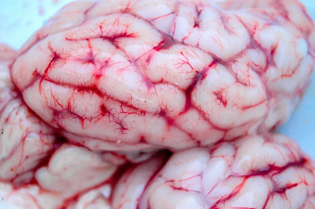
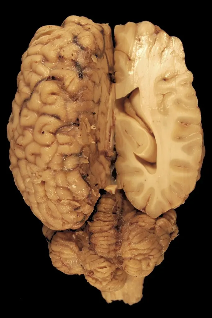
Laminar necrosis is the cortical pattern of this injury. The chapter notes that cortical damage sometimes causes a band-like lesion, called pseudolaminar necrosis. The word “sometimes” matters: not every case of global ischemia produces a cortical band, so its absence does not rule out hypoxic-ischemic injury.
Watershed injury is another pattern, recognised on neuroimaging in neonatal hypoxic-ischemic encephalopathy, as described on Pediatric Education. Watershed zones lie between arterial territories. When a cortical band or a watershed pattern is present, still check the hippocampus and cerebellum for neuronal loss, because those are the most consistent vulnerable sites.
Global ischemia compared with focal infarct
Global ischemia affects the whole brain, usually after cardiac arrest or severe hypotension (shock). A focal cerebral infarct is confined to the territory of one disturbed vessel. The practical difference is distribution: global injury follows cell vulnerability, so scattered neuron populations suffer, while a focal infarct follows the vascular territory.
The chapter also ranks insults. Global ischemia is worse than hypoxia, hypoglycemia and seizures because it adds lactic acid and toxic metabolite accumulation. Sustained severe hypoglycemia, and seizures lasting 1-2 hours, also cause permanent damage, so a history containing those events does not make a purely ischemic explanation the only possibility.
In the developing brain, the pattern depends on the age at insult. In the rat models described in PubMed Central, hypoxia-ischemia in the youngest pups produces multifocal white-matter lesions, while insult at later postnatal ages produces severe parasagittal cortico-subcortical infarcts. In that model, apoptotic cells also increased in offspring after 10 and 15 minutes of perinatal hypoxia. These are animal findings, so use them to explain why neonatal patterns differ from adult ones, not as direct measurements in human newborns.
In newborns, HIE occurs in 1-3 per 1000 live births, and acute catastrophic asphyxia is associated with about 25% of cases, according to Pediatric Education. Infants with moderate to severe HIE have a 48% chance of major neurological problems or death even with hypothermic treatment. When severe swelling accompanies an injury, the mechanical consequences are covered under brain herniation types.
Why neurons are vulnerable
Energy demand explains much of the selectivity. The brain is about 2% of body mass but consumes 15% of the energy generated in the body, according to the neuropathology-web.org chapter. When perfusion fails, the tissue has little reserve.
The chapter gives two reasons why neurons are more sensitive than glial cells: they have higher energy demands, and only they produce glutamate. Selective vulnerability follows from this, since the neuron populations listed above are the ones that fail within the shortest ischemic window.
Global ischemia is also more damaging than hypoxia alone because it adds lactic acid and toxic metabolite accumulation. That is why a patient with a period of absent perfusion often has more extensive neuronal loss than one with low oxygen but preserved flow.
Frequently asked questions
How soon do hypoxic-ischemic changes appear under the microscope?
They begin within hours of restored perfusion, and some neurons die hours or days later, according to the neuropathology-web.org chapter.
What usually causes global ischemia?
Cardiac arrest or severe hypotension (shock) is the usual cause. Focal ischemia, by contrast, produces an infarct in one vascular territory.
How often does HIE occur in newborns?
It occurs in 1-3 per 1000 live births, according to Pediatric Education.
Leave a Reply