
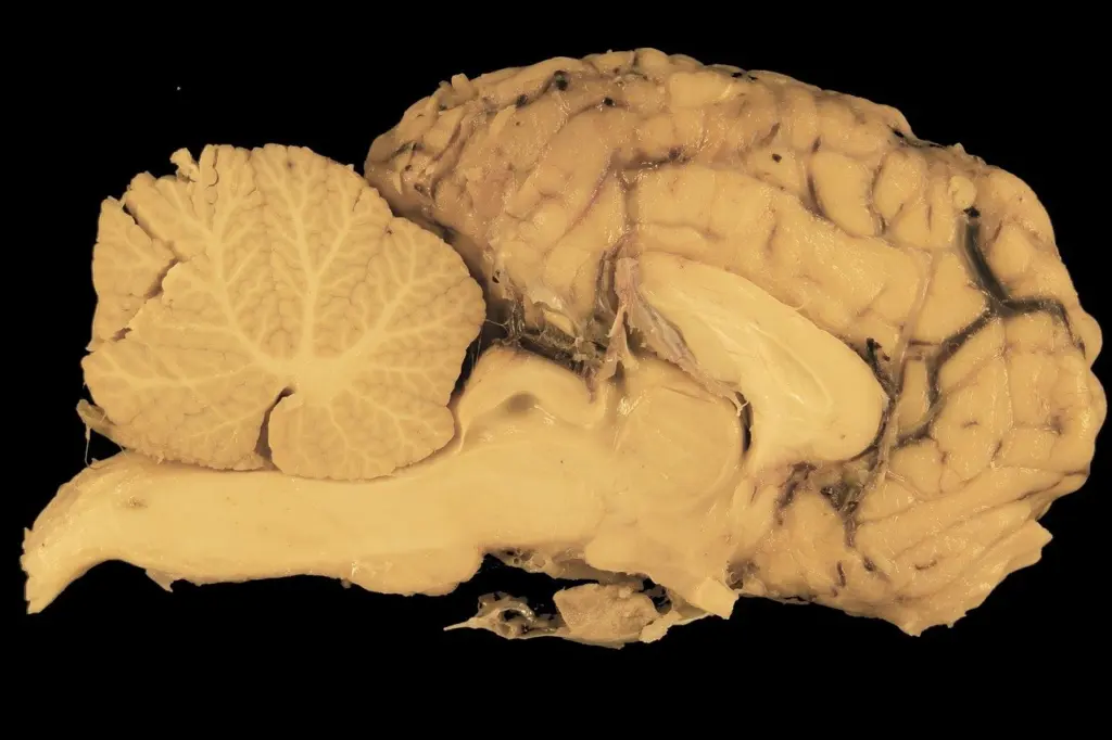
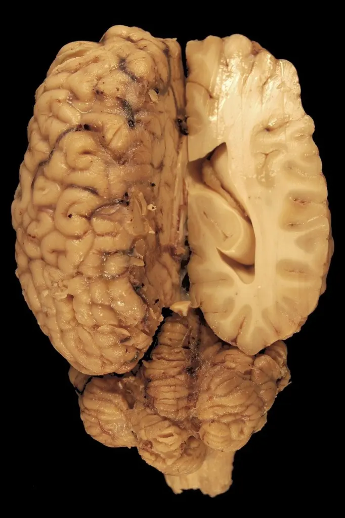
A swollen brain at autopsy with a grooved uncus or a displaced cingulate gyrus shows the main brain herniation types at work: five are described, and each compresses its own structures, so gross findings predict clinical signs.
This page is part of the neuropathology course. It links edema mechanisms to herniation patterns, then shows how each pattern maps onto damaged structures and expected signs.
Why swelling leads to herniation
Herniation follows from a pressure problem inside a closed box. The Monro-Kellie principle, as set out in a European Society of Radiology teaching poster, states that the sum of the volumes of brain, cerebrospinal fluid and intracranial blood is constant in an intact skull. When one compartment grows, the others must shrink or tissue must move. Once compensation fails, the brain is pushed across the rigid dural folds and bony openings that divide the cranial cavity.
The same ESR poster sorts the causes of raised intracranial pressure by compartment:
- Brain tissue and expansive lesions: a mass adds volume to the brain compartment directly.
- Brain water content: cytotoxic and vasogenic edema both increase the intracerebral water content, and so both enlarge the brain compartment.
- Venous blood: venous obstruction raises the intracranial blood volume.
- Cerebrospinal fluid: hydrocephalus enlarges the fluid compartment.
Cytotoxic and vasogenic edema are therefore two routes to the same pressure outcome. For herniation, the practical question is usually how much volume has been added and where, not which edema type produced it. Hypoxic injury, a common setting for brain swelling, is covered in hypoxic ischemic encephalopathy neuropathology.
Herniation types compared
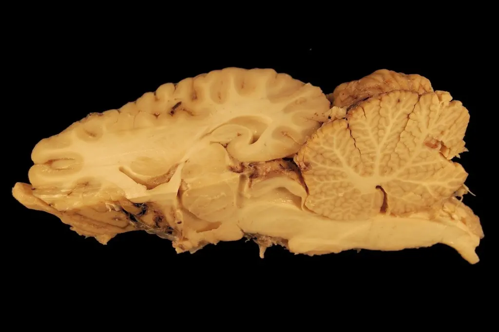
The ESR poster describes five varieties of cerebral herniation: subfalcine, transtentorial, tonsillar, transphenoidal and transcalvarial. Subfalcine herniation, in which the cingulate gyrus is displaced under the falx cerebri according to a Lecturio clinical summary, is the pattern met most often. The transtentorial pattern is usually discussed through its uncal form. The table covers the three patterns with the most distinctive structural consequences.
| Type | Structures compressed | Expected consequence | Planning use |
|---|---|---|---|
| Subfalcine | Ipsilateral lateral ventricle and foramina of Monro; the ICA and ACA can buckle with significant shift | Obstructive hydrocephalus with continued mass effect; possible arterial occlusion | Check ventricular size and midline shift first |
| Uncal (descending transtentorial) | Suprasellar cistern, cerebral peduncle and oculomotor nerve; the posterior cerebral artery can also be compressed | Pupillary dilation and loss of light reflex; ischemia of the ipsilateral primary visual cortex with contralateral homonymous hemianopia | Look for uncal grooving and cistern effacement |
| Tonsillar | Cervicomedullary core and central canal; PICA at the foramen magnum margins | Blocked central canal (hydromyelia); possible PICA compression | Inspect the cerebellar tonsils against the foramen magnum |
Attribution: structures and consequences for subfalcine and uncal herniation come from the ESR poster, and arterial and tonsillar details from the University of Florida neuroradiology guide.
Direction matters for transtentorial herniation
The University of Florida guide separates downward from upward transtentorial herniation by the cisterns involved. Downward herniation displaces predominantly ventral structures and affects the suprasellar and ventral circummesencephalic cisterns. Upward herniation affects the dorsal incisural cisterns. When a specimen or scan shows effacement of the ventral cisterns, think downward descent of the uncus rather than an upward push from the posterior fossa.
Common misunderstandings about herniation patterns
Several assumptions trip up students who learn herniation as a list of names rather than a set of structural relationships.
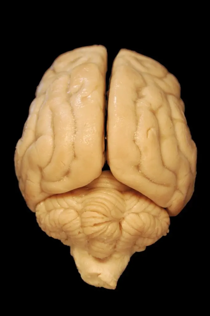
- “Subfalcine herniation is the minor one.” It is the most common type, but frequency is not severity. The ESR poster reports that a shift of the midline greater than 15 mm correlates with a poor prognosis, while a shift under 5 mm does not affect outcome. Treat the degree of shift as the finding that matters, and be wary of calling any subfalcine displacement trivial without measuring it.
- “Uncal herniation always paralyses the opposite side.” Often it does not. The uncus may press the contralateral cerebral peduncle against the tentorium cerebelli, producing ipsilateral hemiparesis. This false localizing sign is the reason the grooved notch on the opposite peduncle matters at autopsy. A student who reads weakness side alone will point to the wrong hemisphere.
- “The pupil is a late curiosity.” Pupillary dilation with loss of the light reflex follows directly from compression of the oculomotor nerve, which carries parasympathetic innervation. It is a structural consequence of the uncus crossing the tentorial edge, not a vague warning sign.
- “Tonsillar herniation and Chiari malformation are the same thing.” They overlap in what is displaced, but the University of Florida guide notes that Chiari malformations have tonsillar herniation often compressing the cervicomedullary cord without displacement at the tentorial incisura. Secondary tonsillar descent from raised pressure belongs to a broader pattern of intracranial displacement.
- “Compression means only nerve and tissue damage.” Arteries are vulnerable too. Significant subfalcine shift can buckle the ICA and ACA, uncal herniation can compress the posterior cerebral artery, and downward tonsillar herniation can compress PICA where it arises near the C1 level. Check vessel territories as well as parenchyma when you reason from gross findings.
Duret hemorrhage in the brainstem
Duret hemorrhages are secondary hemorrhagic infarcts of the midbrain that follow brainstem compression. The ESR poster explains the mechanism: perforating arteries are compressed and buckled as the brainstem is displaced, producing a secondary hemorrhagic midbrain infarct known as a Duret hemorrhage. They are a consequence of herniation, not a cause of it, which is why they are found at autopsy in the central midbrain of patients with marked downward displacement.
There is a second route to the same lesion. The University of Florida guide notes that when mass effect resolves, reperfusion hemorrhage can develop in the central mesencephalon. A Duret hemorrhage therefore does not always mean compression was ongoing at the moment of death. It can also reflect blood returning to vessels that were squeezed. For a student, the lesion marks the brainstem as the site where herniation turned from a displacement into irreversible tissue injury.
Reading gross findings to predict signs
The most useful habit is to work from structure to sign. Start with the displaced tissue, name the structure it presses on, and the sign follows from that structure’s function.
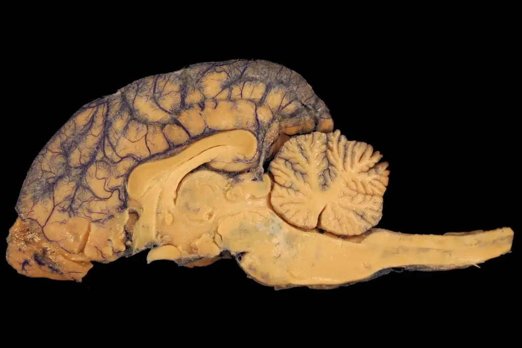
- Identify the displacement. Cingulate gyrus under the falx means subfalcine herniation. A grooved uncus over the tentorial edge means uncal herniation. Tonsils below the foramen magnum plane mean tonsillar herniation.
- Name what lies in the path. For uncal herniation that is the oculomotor nerve, peduncle and posterior cerebral artery. For subfalcine herniation it is the foramina of Monro and anterior cerebral artery. For tonsillar herniation it is the medulla, cervical cord junction and PICA.
- Predict the sign. Oculomotor compression gives a fixed dilated pupil, posterior cerebral artery ischemia gives visual field loss, and foramen of Monro obstruction gives hydrocephalus.
- Check for secondary injury. Midbrain hemorrhage points to Duret hemorrhage, and arterial buckling points to infarcts in the vessel’s territory.
This approach keeps the focus on anatomy rather than rote lists. Seizure-related brain changes follow a different pattern, covered in epilepsy neuropathology.
Frequently asked questions
| Question | Answer |
|---|---|
| Which herniation type is the most common? | Subfalcine herniation, according to the ESR poster, which describes it as the most common brain herniation. |
| What is Cushing’s triad? | A combination of hypertension, bradycardia and irregular breathing that indicates increased intracranial pressure, as summarised by Lecturio. |
| Is brain herniation an emergency? | Yes. UF Health describes it as a medical emergency in which treatment aims to save life by reducing swelling and pressure, using medicines such as mannitol, saline or other diuretics. |
| Can a Duret hemorrhage appear after pressure falls? | It can. Reperfusion hemorrhage into the central mesencephalon has been described once mass effect resolves, per the University of Florida neuroradiology guide. |
Leave a Reply