

Quick answer: Alzheimer’s disease neuropathology is defined by extracellular amyloid plaques and intraneuronal neurofibrillary tangles, read from the gross brain down to the microscope. Both lesions are still required for a pathological diagnosis, and most cases also carry other co-pathologies.
This guide follows the order a student meets the findings at the autopsy bench. For the wider family of disorders it belongs to, see the overview of types of neurodegenerative diseases.
Deciding what to look at first

The sequence that works best at the bench moves from what the naked eye can judge to what only a stain can show. Each step narrows the question, and each lesion has its own protein target.
- Start with the gross brain. Weight and the pattern of gray matter loss tell you whether the brain fits a neurodegenerative process, but they cannot name the disease.
- Move to plaques. Plaques are made of beta-amyloid peptide, so an amyloid beta antibody is the logical target for them.
- Then assess tangles. Tangles are made of hyperphosphorylated tau, so a phospho-tau antibody is the logical target for them.
- Check the vessels. Amyloid can also sit in vessel walls, and it should be judged separately from parenchymal plaques.
- Finish with co-pathologies and incidental changes. Other proteinopathies and age-related processes decide how much of the picture Alzheimer pathology explains.
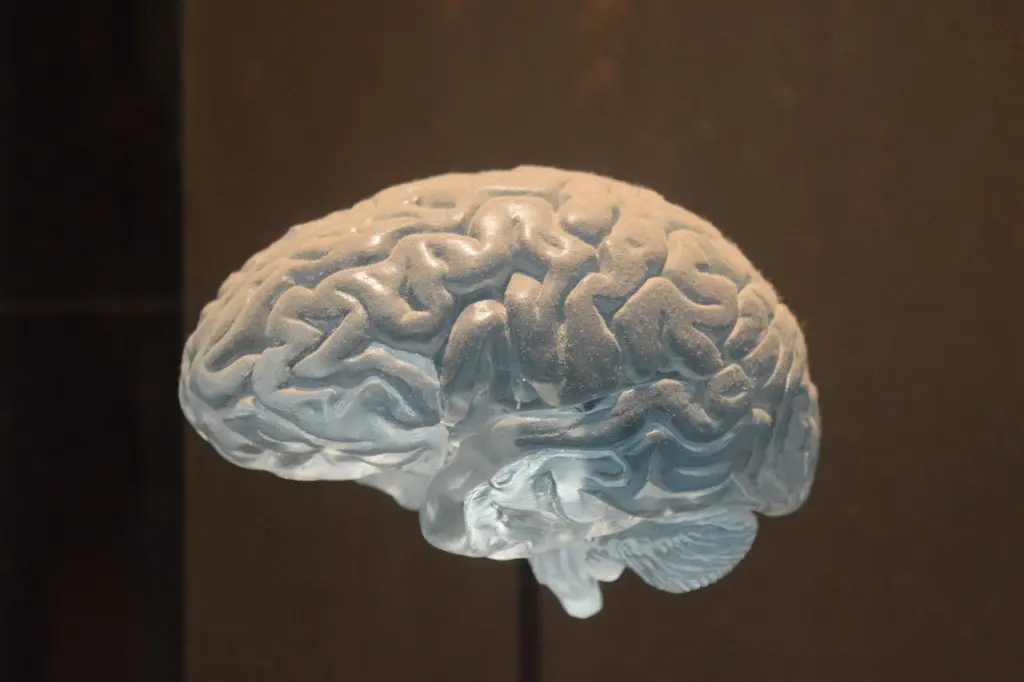
Gross findings: brain weight and gray matter loss
In the Mayo Clinic Brain Bank series, brains with Alzheimer disease and no other major pathology had a measured mean weight, according to the Mayo neuropathological diagnosis review. The wide spread among individual brain weights is the practical lesson. Brain weight on its own cannot confirm or exclude Alzheimer disease, so treat it as supporting context for the microscopy.

Gray matter loss is the gross counterpart of the microscopic story. A Texas A&M review of tau spread notes that the misfolding, spread and accumulation of aggregates precede gray matter loss in the affected regions. When you see atrophy, the lesions that caused it have therefore been present for some time.
Why microscopy decides the diagnosis
Clinical Alzheimer dementia cannot be definitively diagnosed until postmortem neuropathologic evaluation, as the Mayo neuropathological diagnosis review states. A Mayo review of Alzheimer disease and other dementias makes the same point, describing neuropathology as the only way to make a definite diagnosis. A dementia syndrome that looks like Alzheimer disease in life is a clinical label until the tissue has been examined.
Amyloid plaques versus neurofibrillary tangles
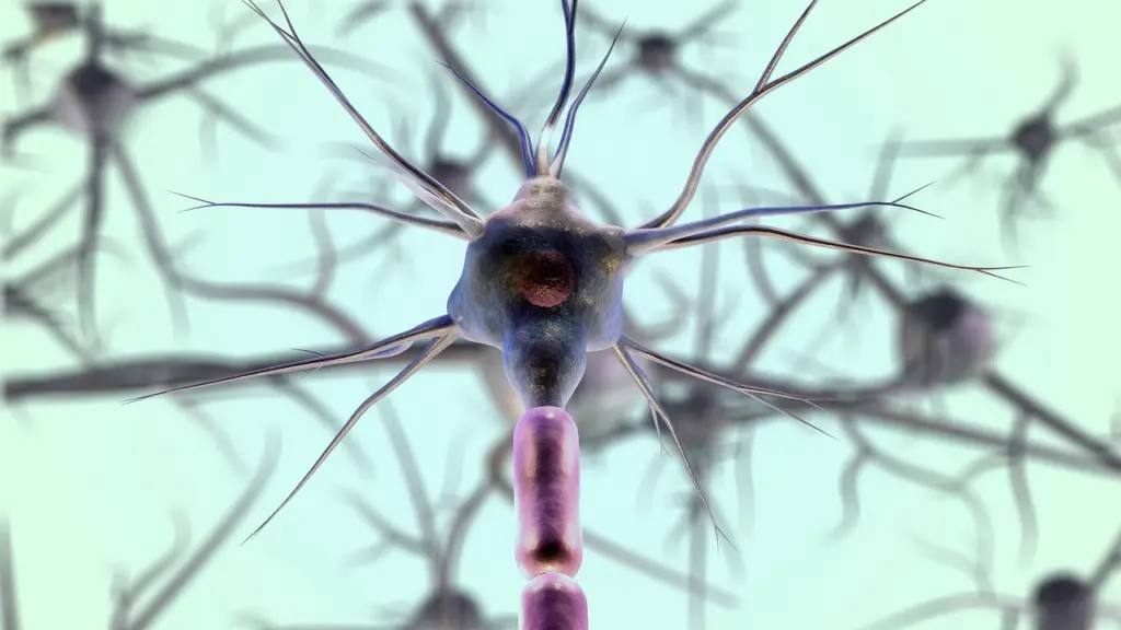
The two defining lesions differ in location and in composition. Amyloid plaques are extracellular deposits of beta-amyloid peptides, according to an overview of the neuropathological hallmarks. Neurofibrillary tangles are intraneuronal aggregates of hyperphosphorylated, misfolded tau. Under the microscope, that means plaques lie between cells, while tangles sit inside neurons and fill the cell body.
Tau becomes a tangle in stages. The Texas A&M review describes tau that has been phosphorylated and misfolded forming insoluble neurofibrillary tangles that aggregate and spread. BrainFacts adds that tangles can kill neurons, which is one reason tangle burden matters beyond simple lesion counting.
| Lesion | Location | Composition | Planning use |
|---|---|---|---|
| Amyloid plaque | Extracellular, in the tissue between cells | Beta-amyloid peptides | Choose an amyloid beta target to show plaque distribution. |
| Neurofibrillary tangle | Inside neurons | Hyperphosphorylated, misfolded tau | Choose a phospho-tau target to show tangle burden within cells. |
| Cerebral amyloid angiopathy | Tunica media of leptomeningeal arteries and cortical capillaries | Beta-amyloid | Assess vessel walls separately from plaques in the parenchyma. |
| Granulovacuolar degeneration and Hirano bodies | Neurons | Not defined by the pack | Record them, but do not weigh them as diagnostic. |
Lesion descriptions in the table come from the neuropathological hallmarks overview and the Mayo diagnostic review.
Where the pathology starts and where it spreads
Pathological tau is shown to spread through the limbic network, and the Texas A&M review compares this pattern to the neuronal spreading proposed by Braak and Braak. That framework is the basis for staging tangle distribution, covered in detail under braak staging. Use staging when you need to describe how far the disease has advanced. Use the lesion types above when you need to say what the disease is.
Because the diagnosis requires both plaques and tangles, tangle changes in an older brain without matching plaques raise a different question. That situation is discussed under primary age-related tauopathy.
Cerebral amyloid angiopathy in Alzheimer disease
Cerebral amyloid angiopathy (CAA) is beta-amyloid deposition in vessel walls, specifically the tunica media of leptomeningeal arteries and cortical capillaries. It is the vascular face of the same amyloid process. The hallmarks overview reports that some level of CAA, often mild, is present in nearly all Alzheimer brains.
Severity is what changes the clinical consequence. Severe CAA may weaken the vessel wall and ultimately lead to lobar hemorrhage. When you grade CAA at the bench, a mild finding is expected, while a severe one deserves explicit mention in the report.
The Mayo series tabulates CAA groups separately within its 1153 patients with a pathologic diagnosis of Alzheimer disease, listing 42 in the AD/CAA group and 110 in the AD/CAA+ group, according to the Mayo neuropathological diagnosis review. Read those counts as group sizes within one bank series. They are not a general prevalence figure.
Mixed pathology and incidental findings
Alzheimer disease is best treated as a mixed proteinopathy of amyloid and tau that is frequently associated with other age-related processes. In the Mayo Clinic Brain Bank, it is rarely found without other neurodegenerative co-pathologies. Across the clinical Alzheimer cases the Mayo review examined for 2007 to 2016, the majority were found to have co-pathologies. A report that lists only plaques and tangles is therefore usually incomplete.
Granulovacuolar degeneration and Hirano bodies belong in this section too. The same review calls them inevitable findings of uncertain significance. They are common enough to expect and worth describing, but they should not drive the diagnosis.
Troubleshooting common reading errors
- Calling a brain Alzheimer disease from weight alone. The mean weight of 1077 g carries a spread of 159 g, so many brains will overlap. Confirm with plaques and tangles.
- Expecting a pure case. If you find only amyloid and tau, check again for co-pathologies before concluding that nothing else is present.
- Over-reading secondary changes. Granulovacuolar degeneration and Hirano bodies support a neurodegenerative context but do not establish the diagnosis.
- Ignoring vessel walls. A section that looks clear of plaques can still show amyloid in the vessel wall. Examine leptomeningeal and cortical vessels deliberately.
- Confusing the lesions. If a deposit lies between cells, think plaque. If it fills the neuron, think tangle.
Frequently asked questions
How much of dementia is Alzheimer disease?
Alzheimer disease is the most common form of dementia, accounting for 60 to 80% of cases according to the Mayo neuropathological diagnosis review.
Do neurofibrillary tangles damage neurons?
Yes. BrainFacts notes that tangles can kill neurons, which is why tau pathology is treated as more than a passive marker.
Who first described the disease, and which features still define it?
Alois Alzheimer described it more than 100 years ago. The cardinal features he described, amyloid plaques and neurofibrillary tangles, are still required for pathological diagnosis.
Is cerebral amyloid angiopathy a separate disease?
It is a distinct vascular lesion, but it is commonly found alongside Alzheimer pathology. Report it as its own finding and grade its severity.
Leave a Reply