
| Disease or group | Abnormal protein or lesion | Pattern of cell loss on examination | Planning use |
|---|---|---|---|
| Alzheimer disease | Amyloid beta plaques and tau tangles | Cortical atrophy with enlarged ventricles; swollen axons near plaques on electron microscopy | Start here to learn the plaque and tangle vocabulary that other tauopathies are measured against |
| Parkinson disease and Lewy body disease | Alpha-synuclein in Lewy bodies | Loss of pigmented neurons, seen grossly as a pale substantia nigra | Start here for the synucleinopathy side of the comparison |
| Frontotemporal dementia | Tau or another protein, depending on the subtype | Frontal and temporal lobe atrophy | Use it to learn why one clinical syndrome can have different proteins underneath |
| Amyotrophic lateral sclerosis | protein inclusions in motor neurons; a repeat expansion in about half of inherited ALS and frontotemporal dementia cases | Motor neuron loss, with pallor of the corticospinal tract in the spinal cord | Read it next to frontotemporal dementia, because the two overlap genetically |
| Huntington disease | Polyglutamine buildup from a CAG repeat | Caudate nucleus atrophy | The cleanest example of a single inherited cause producing a recognisable gross pattern |
The hallmark lesions, the repeat expansions and the swollen-axon finding above follow Yale Medicine’s overview of neurodegenerative disease mechanisms, and the shared neuronal loss follows a peer-reviewed review on neurodegeneration.
The main types of neurodegenerative diseases are easiest to compare by the abnormal protein that accumulates and the neurons that die. Alzheimer, Parkinson, frontotemporal dementia, ALS and Huntington disease are the core examples.
This page is a map for students of neuropathology. Begin with the shared features and the microscope findings, then follow whichever disease family matches your course. Each family links to a fuller guide, for example alzheimer’s disease neuropathology for the most common dementia.
Main neurodegenerative diseases and what unites them
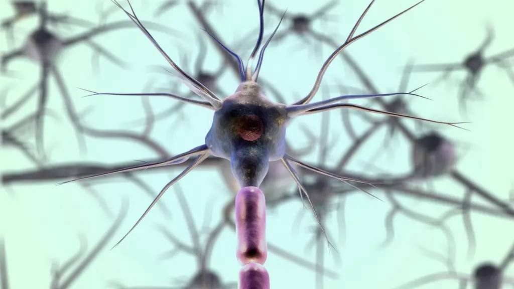
Neurodegenerative diseases are heterogeneous neurological disorders characterised by a progressive loss of selected neuronal populations, according to a review indexed in PubMed Central. The word selected matters. Each disease attacks particular cell types in particular regions, and that selectivity is what makes the diseases distinguishable at autopsy.
Shared features
The same review notes that, despite different aetiologies and symptoms, all of these diseases feature progressive loss of selected neurons. BrightFocus describes neurodegeneration in plain terms: neurons in the brain or peripheral nervous system lose function over time and ultimately die. For a student, this gives a useful first rule. When a case shows cell loss, the next question is not whether neurons died but which ones, and in what distribution.
The adult-onset group named in the review includes Alzheimer disease, Parkinson disease, Huntington disease and amyotrophic lateral sclerosis. Frontotemporal dementia belongs alongside them, and it shares genetics with ALS, as the protein section below shows. Many diseases in this broad group are characterised by proteins that fold incorrectly and pile up as aggregates in the brain, as Yale Medicine explains.

Which diseases are most common
BrightFocus, citing the National Institute of Environmental Health Sciences, reports that Alzheimer disease and Parkinson disease are the most common neurodegenerative diseases. That ordering is a good guide to how much teaching time each deserves. Alzheimer and Parkinson disease are the two to master first, because most of the other diseases are described by contrast with them. Huntington disease, ALS and frontotemporal dementia matter for their teaching value, since each shows a distinct mechanism or a distinct pattern of loss.

Age is the other organising fact. A significant risk factor for most of these diseases is ageing, and Lee Health’s neurology pages state that the risk rises dramatically with age. This explains why age-related pathology that falls short of disease, such as primary age-related tauopathy, is a constant complication when you read an older person’s brain.

What neurodegeneration looks like under the microscope
Neurodegeneration under the microscope is, at its simplest, a shortage of neurons in a region where they should be abundant, plus the debris and reactive changes that follow. The review cited above traces early evidence to neuropathological observations of disrupted microtubule arrangement and dystrophic neurites. Those are the structural signs that a neuron’s internal transport and architecture have failed before the cell is lost.
Gross findings
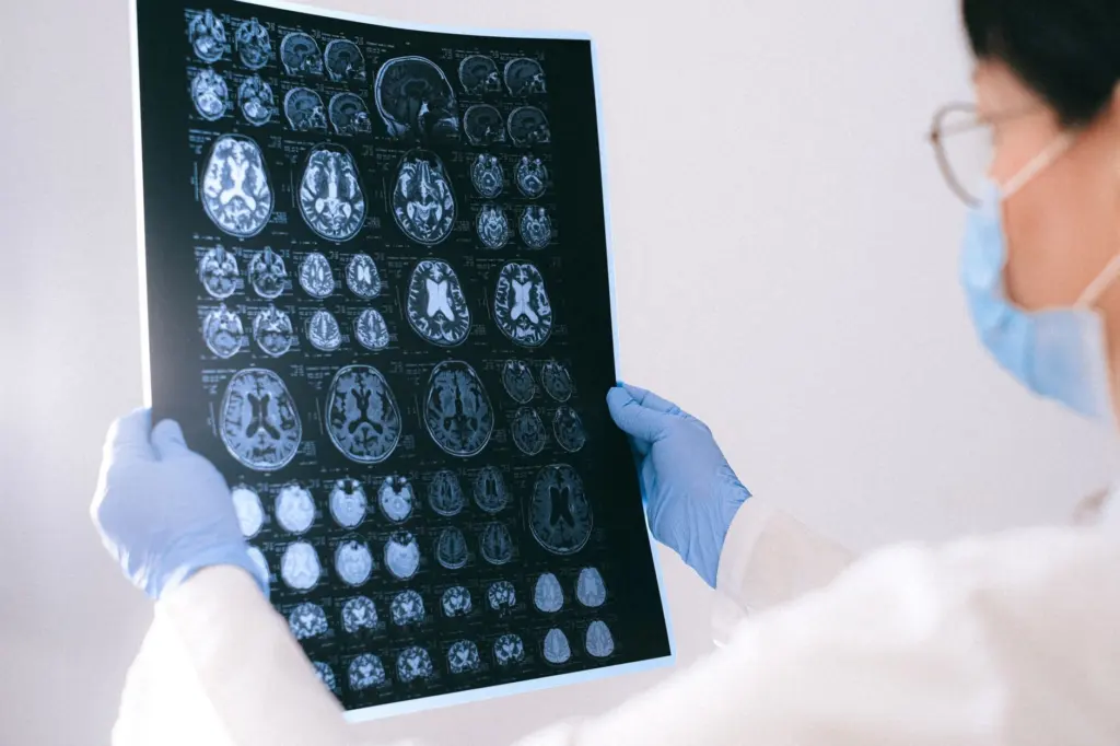
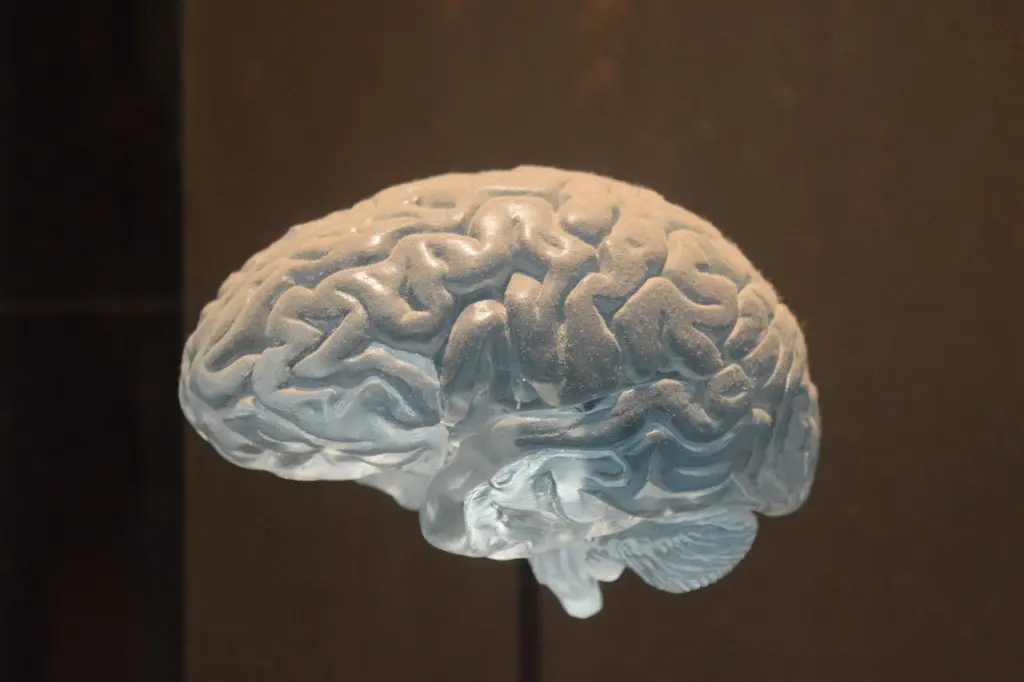
At the cut brain, the key gross finding is atrophy. Alzheimer disease classically shows shrunken cortex with enlarged ventricles, because lost tissue is replaced by space. Parkinson disease shows its loss in the brainstem, where the substantia nigra loses pigment as pigmented neurons die. Huntington disease shows its loss deep in the brain, in a shrunken caudate nucleus. Frontotemporal dementia concentrates its loss in the frontal and temporal lobes, while ALS shows its main change in the spinal cord, where corticospinal tract degeneration appears as pallor.
The practical lesson is to read the location before the lesion. A pattern of atrophy narrows the differential before any stain is applied, and it tells you where to sample.
Microscopic findings
Under the microscope, you look for neuron loss, gliosis, and the protein deposits that characterise the disease. Electron microscope images of Alzheimer brain show clusters of swollen axons near amyloid plaques, which Yale Medicine describes as a hallmark of the disease. Impaired axonal transport has been reported in Alzheimer disease, ALS and Parkinson disease alike, according to the PubMed Central review. That makes transport failure a shared mechanism worth knowing, but not a distinguishing feature, since it appears in several diseases.
Neurons make this process hard to reverse. Yale Medicine notes that neurons are largely irreplaceable and must function for nearly a century. A region that has lost its neurons stays depleted, so the severity of loss you see at autopsy is cumulative over the whole course of the illness.
Proteins that accumulate in different diseases
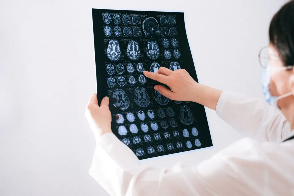
The most useful way to organise these diseases is by the protein that accumulates, because the protein predicts the inclusion, the distribution and the name of the disease family. Yale Medicine’s overview describes the general mechanism: proteins fold incorrectly and then pile up as aggregates in the brain.
Tauopathies versus synucleinopathies
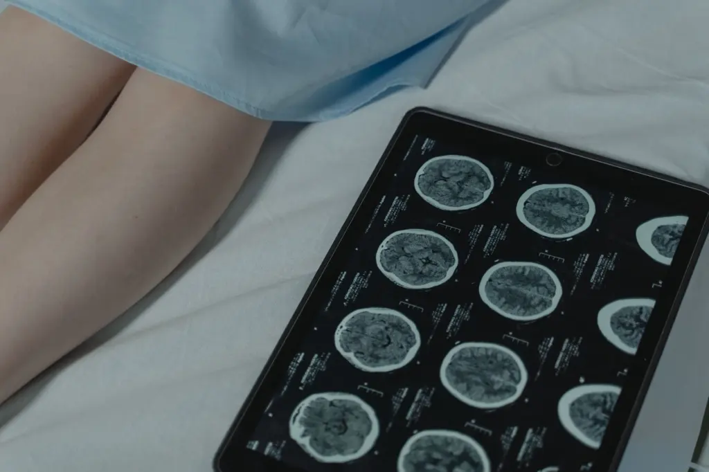
A tauopathy is a disease in which the tau protein accumulates abnormally in neurons or glia. A synucleinopathy is a disease in which alpha-synuclein accumulates instead. The difference is therefore the identity of the protein, and it decides which immunostain you reach for. Tau-based diseases include Alzheimer disease, pick disease pathology, progressive supranuclear palsy pathology and chronic traumatic encephalopathy pathology. Synuclein-based diseases include Parkinson disease, dementia with Lewy bodies and multiple system atrophy.
Do not treat the two groups as mutually exclusive in every brain. Alzheimer-type changes and Lewy-type changes frequently coexist in older people, so a single case can carry both labels. Naming the dominant protein first, then checking for the second, is the safer routine.
Amyloid beta and tau in Alzheimer disease
Alzheimer disease is unusual because two proteins are involved. Amyloid beta accumulates as plaques, and tau accumulates as neurofibrillary tangles inside neurons. Yale Medicine reports that amyloid begins piling up as much as 10 years before people begin developing symptoms. The same overview describes a cascade in which amyloid beta, through its association with prion protein, activates a receptor that recruits inflammatory mediators and tau to the synapse. The clinical consequence is that plaques tell you about the disease early, while the tangles are the pattern you stage against, as the autopsy section below explains.
Shared protein inclusions in ALS and frontotemporal dementia
ALS and frontotemporal dementia are often grouped because they share a molecular background. Yale Medicine reports that about half of all inherited cases of ALS and frontotemporal dementia involve a repeating sequence. At the protein level, much of frontotemporal lobar degeneration and most ALS involve protein inclusions, which is why a guide to TDP-43 proteinopathy is the natural companion to either disease. Some frontotemporal cases are tau-based instead, so this family is the best example of one clinical label with more than one protein behind it.
Polyglutamine in Huntington disease
Huntington disease is the textbook case of a repeat disorder. According to Yale Medicine, the repeating nucleotide triplet CAG causes the polyglutamine buildup in the disease. It differs from Alzheimer disease in that a single inherited repeat causes it. By contrast, BrightFocus notes that late-onset Alzheimer disease cannot be inherited as a single mutation that on its own causes the disease.
Alzheimer disease and the tauopathy family
Start with Alzheimer disease if you want the reference pattern. It combines the most recognisable gross change, cortical atrophy, with the two best-known microscopic lesions. Plaques and tangles are what every other tau-based disease is compared with, so learning them well saves effort later.
Choosing the next tauopathy
- Learn the staging logic first. The tangle distribution is organised by braak staging, which is the backbone of Alzheimer neuropathologic assessment and the reason regional sampling matters.
- Move to Pick disease for a contrasting tau pattern. It is a frontotemporal disease with rounded tau inclusions, a useful contrast with tangles.
- Choose progressive supranuclear palsy for brainstem-predominant tau. It shows how a tauopathy can be defined by region rather than by tangles in the cortex.
- Add argyrophilic grain disease for a subtle tau pattern. Argyrophilic grain disease is a good lesson in why silver stains and immunostains reveal pathology that the haematoxylin and eosin stain misses.
- Use primary age-related tauopathy as a boundary case. It teaches you where ageing ends and disease begins.
- Save chronic traumatic encephalopathy for the exposure link. It shows a tauopathy tied to repetitive head injury, with its own distribution.
If you want one reference to hold the whole family together, the list of tauopathies sorts the diseases by tau isoform and cell type. That makes it the efficient overview once you know two or three members well.
Parkinson disease and the synucleinopathies
Parkinson disease is the second anchor disease. Its gross signature is the pale substantia nigra, and its microscopic signature is the Lewy body in a surviving pigmented neuron. Both findings are the same event seen at different scales, since the pigment is lost when the neurons that carry it die.
Parkinson disease and Lewy bodies
For a full account of the brainstem findings, read the neuropathology of parkinson’s disease. For the inclusion itself, with its stains and its spread, read alpha-synuclein pathology. Take them in that order, because the gross picture gives context for the cellular one.
Related synucleinopathies
Two diseases extend the synuclein story. Dementia with lewy bodies pathology reaches the cortex, which is why it presents with cognitive decline rather than movement problems alone. Multiple system atrophy pathology places the same protein in glial cells instead of neurons. This difference in the cell type involved is the main reason it is taught as a separate disease.
ALS, frontotemporal dementia and the motor-cognitive overlap
Study these diseases together. ALS selectively destroys motor neurons, producing corticospinal tract pallor in the spinal cord, while frontotemporal dementia destroys frontal and temporal cortex. The shared repeat expansion described above is the clearest evidence that they are related, and the shared protein inclusions are what unite many of the cases.
A sensible route is to read about the cortical disease first and then the spinal disease, because the cortical guide explains the subtypes. The overview of frontotemporal lobar degeneration pathology separates the tau subtypes from the non-tau subtypes. That separation is the key to the whole group, since the same clinical picture can carry different pathology. A student who has seen both subtypes will not assume that a frontotemporal syndrome means a tauopathy.
Huntington disease
Huntington disease teaches a different lesson from the protein-aggregate diseases above, since its cause is a known inherited repeat. The gross result is atrophy of the caudate nucleus, and the cellular result is the loss of a particular neuron population. For the staging, the inclusions and the gross pattern, use the guide to huntington’s disease pathology.
Choose this disease early if your course emphasises genetics. Choose it later if your course emphasises dementia, because Huntington disease is rarely the diagnosis in a memory-clinic case.
How a neurodegenerative disease is confirmed at autopsy
Confirmation at autopsy rests on tissue. A neuropathologist examines the whole brain for the pattern of atrophy, samples defined regions, and applies stains that highlight the protein deposits. The diagnosis is then made by matching the distribution and type of deposits against the published criteria for each disease. A clinical diagnosis during life is a working label, and the tissue is what confirms or revises it.
Why tissue and not genetics
Genetic information does not replace the examination. BrightFocus notes that late-onset Alzheimer disease cannot be inherited as a single mutation that alone causes the disease, so there is no simple genetic test that settles the diagnosis. For Huntington disease the inherited repeat is the cause, yet the tissue examination still documents the extent of loss. In practice, the neuropathologist reports the dominant protein, the regional distribution and any coexisting pathology, such as Alzheimer-type changes in a case labelled as Parkinson disease.
Staging and criteria
Staging systems turn a pattern into a reproducible grade. For Alzheimer disease, tangle distribution and plaque burden are combined in the criteria discussed in the Braak staging guide. The principle carries over to the other diseases: sample consistently, stain for the right protein, and report distribution rather than just presence. Without regional sampling, a mild or early case can be missed or misclassified.
Prevalence, risk and the limits of treatment
The National Institute of Environmental Health Sciences, as cited by BrightFocus, names Alzheimer and Parkinson as the commonest neurodegenerative conditions. For the rest, treat prevalence as a reason to prioritise your reading rather than as a figure to memorise. Ageing raises risk across most of the group, which is why the same brain often carries more than one pathology.
Treatment sets the clinical context for the pathology. Lee Health states that there is no definitive cure for most of these complex neurological diseases. That is the reason the pathologic diagnosis carries so much weight: it defines what the disease was, even when the clinical course could not be changed.
Where to start, by course goal
- General neuropathology course. Read the glance table, then the microscopy section, then Alzheimer and Parkinson disease in that order.
- Dementia focus. Work through Alzheimer disease, frontotemporal lobar degeneration, dementia with Lewy bodies and the tauopathy list.
- Movement disorder focus. Begin with Parkinson disease and alpha-synuclein pathology, then add multiple system atrophy and progressive supranuclear palsy.
- Motor neuron and genetics focus. Read ALS and protein inclusions together, then Huntington disease as the contrasting inherited example.
- Exam preparation. Rehearse the comparison method: name the protein, name the region, name the gross pattern, and check for a coexisting pathology.
Each route works because the protein, the region and the gross pattern are consistent within a disease and different between diseases. That consistency is the reason the unified approach in this guide is more efficient than memorising disease descriptions one by one.
Frequently asked questions
Are all neurodegenerative diseases caused by misfolded proteins?
Not all of them, but many are. Yale Medicine describes many neurodegenerative diseases as characterised by proteins that fold incorrectly and aggregate in the brain. Huntington disease is a good example of a different route, since its polyglutamine buildup follows from an inherited CAG repeat.
Can the brain replace neurons lost to these diseases?
No. BrightFocus notes that, unlike skin, muscle and bone cells, neurons do not regenerate. Lost neurons stay lost, so the pathology seen at autopsy reflects loss accumulated over the illness.
Do neurodegenerative diseases occur in children?
Yes. Lee Health lists neuronal ceroid lipofuscinosis and Werdnig-Hoffmann disease among pediatric neurodegenerative types. These differ from the adult-onset diseases covered here, which include Alzheimer, Parkinson, Huntington and ALS.
Is there a cure for neurodegenerative diseases?
For most of them, there is no definitive cure, according to Lee Health. This is why accurate pathologic classification remains central to understanding each disease.
Leave a Reply